Fundus images explained by an ophthalmologist

Vložit
- čas přidán 27. 07. 2024
- FREE FREE FREE !!! FIGURE1 medical app: Discover medical cases from every specialty their views and advice DOWNLOAD NOW download.figure1.com/greenglobe
Prepare for USMLE,UK,CANADIAN,AUSTRALIAN, NURSING & OTHER MEDICAL BOARD examinations around the globe with us.Understand the basics, concepts and how to answer wisely and score 99 in each step. we are here to help you. What are you waiting for subscribe now!!!
SUBSCRIBE NOW: bit.ly/161OmbF
For Business inquiries: allornonelaw4business@gmail.com
Join our USMLE step 1 prep Zone : / 730000020375744
Join our USMLE CK STUDY GROUP: / 320959178079398


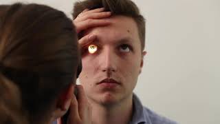


After pupillary dilatation(Tropicamide or cyclopentolate ± phenylephrine):
1.1:05,3:32normal fundus(eye right side)
2.2:55drusens (ARMD)
3.4:15Neovascularisation at optic disc(NVD)
4.4:55,8:12Dot-blot hemorrhages &
microaneurysms
8:48flame-shaped hemorrhage
5.6:01,11:39Hard exudates, laser scars(also 11:06)
6.6:45,7:49,10:18Soft exudates=cotton wool spots
7.9:28diff bw collaterals,new blood vessels
I'm a veterinarian. Recently injured my right eye, and developed glaucoma which is still under treatment. The medication dispensed had develops my interest to study ocular pathology. This is a very nice video for me as I have zero knowledge about Opthalmology. Thank you so much.
Thank you for an excellent presentation. Much appreciated
great presentation - just reviewing knowledge and really helped with the visualisation of what i am seeing and explanations
This is FANTASTIC. Thank you so much for making this, and for posting it, it's so informative,
Great presentation. Thankee
Thanks for selfless teaching. God bless
thanks. you make it easy
Good video but do u have more pathologies? I just could appreciate dm and vein occlusions. any explanations for glaucoma retinal detachments etc?
Very nice.. With medium speed of explanation, so that we can follow... Very informative and practical..
Very useful. thanks.
thank u so much, v helpful..
This is a great educational presentation.
Great vedio👍
Very useful Thankyou 🙏
Great
Good basic examples 👍
Very nice and useful video…thank you
Thank you so much. plz do share our videos.
Thanks.keep it up
Sir is this fundus test find out the treatment of PRK when we takes PRK in recruitment process
This RN who loves A&P, enjoyed your beautiful explanation. Thank You.
Can contura vision Lasik detected by fundas test
Please inform -- I had this done at my eye doctor but she did it several times (with several flashes obviously) on the same left eye, which showed retinopathy. I had the same exam a few days later at the opthamologist (but with just one photo). Now, when there's bright sunshine, I have real vision problems in my left eye.
My question is -- Is there a limit to how many attempts you should make with these cameras on the same eye? -- thanks for any help
Excellent explanation. Thank you so much for posting this
Thanks 👍
Very nice presentation 🙏
Great video!! So useful
Thanks please do share our videos and subscribe.
Standing ovation 😇😇😇
Plz explain flurescein angiography
Great video sir
Great video sir ❤
very nice
Can you guys please let me know what will be reason for an blue dots in fundus photography. I'm an budding r&d specialist and working in developing fundus photography but I came across with this type of error...
Video is very informative, thank you.
but subtitle(scientific words) file have spelling mistakes, please change it.
Kya oct, fundoscopy & ultrasound se retina ke sabhi pblm ka pta chalta h
Be fundus WNL mins?
Doctors Please you help me
I need Good madecin for eyes
Dubil vison
Drops. Serip .Tablit?
Amir Mukhtar Wtf
Very nice sir
Excellent
Wo left eye thaa ray
If my eye pressure is 19 in both eyes with a field vision of 40/40 but my cup disk ratio is about 0.7 in both eyes does that put me at risk for glaucoma? Besides that the doctor did not find any abnormalities.
Your age?
32
thank you!
Any treatment optic nerve please, i am 27 years old. Bike accident 20/3/20 please any treatment my left eye optic nerve is damage
There is an indian on youtube to teach you anything you didnt learn in your life.
😂😂😂
the other day I awoke with my day seem dark as 40 watt light bulb . It sorta like sunset all the time now or wearing sunglasses . like look at old color photographs of 30 and 40 years. I doubt it comes back so sudden.
Think it high blood presures caused .
This is the right eye right
Heard at 2x .!! Good..!!
After Fundus examination, I was super-sensitive to light and near vision was out of focus. Strange feeling !
That is normal following mydriatic drops which disable the iris sphincter muscles and dilate the pupil - this lets in more light as the pupil cannot contract and fine tuning provided by those muscles is also disabled. You should have been warned about this. Shouldn't last more than a few hours.
@@wolf5370 Yes, I was warned about this. It lasted for around 6 hours.
Great effort, thank you very much,,,but slight problem with the accent,pure Indian accent would be better
shri hari He's nri of course he won't have a thick accent.
ay yo safewee
lit vid fam you got me and my mandem bare gassed blad
good nowledge but i couldnt even watch till end cuz the pauses were getting on my nerves
You can try and use 1,5 speed for it, that is what I did :D
There should be captions for this. Can’t understand anything he’s saying. At least I’m not getting captions to show…
Bayonetting sign... ??
debasish panda good question. But, he can't cover everything in just one video. Btw it occurs due to stripping(thinning out) of neuroretinal rim by the increasing siz of cup. Leading to double bending of vessels at the optic disc.
The picture resolution could have been better. Also plz explain all scientific terms u used in video for general population
Hi Mr Opthalmologist, Personally I Think We Should Have Eye Drops With Warfarin Available That Can Be of Great Help..
Don't You Agree?
Thanks
So many ads!😢
I don't understand goodly...
If u an Indian ...can u explained in Hindi ....it's very important and urgent for me...
And I have a question ...my uncle have headache with eye pain last two months ...
I also doing MRI for him.. doctor seen MRI report and saying fundus exam ...for eye....
Sir please help me
Thank you
अगर हिन्दी में दे तो अच्छा जहै
So boring
Nice try.but can definitely be better. 1.Except the first slide the rest of pictures are too small. Whats the need of a large background and a small picture. 2.Very boring abd slow start.go directly to the topic.3.this is a video you make abd later upload so why do you have repeated pictures??abd worst is your comment saying "i think we saw this picture before"!!! Ya we did you have put it again.if its a mistake why don't you correct it before uploading .4.liked the short summary of how to present and what to talk about.but you didnt say anything about how macular edema or other problems look like. I gave a dislike and would give 4 out of 10. Because you can improve and you haven't tried doing so.