Anatomy of the Brain on MRI

Vložit
- čas přidán 20. 10. 2022
- To book a class, come to my website: www.alisanatomycourse.com
This video demonstrates the anatomy of the brain on MRI. It continues with a live interactive anatomical quiz and then to a discussion on common pathological findings.
The Brain
The brain stem
The medulla
The pons
The midbrain
The cerebral peduncles
The cerebellum
The vermis
The left and right hemispheres
Anterior and posterior lobes
The cerebellar tonsils
The cerebral hemispheres
The lobes of the cerebral hemispheres
The frontal lobe
The parietal lobe
The occipital lobe
The temporal lobe
The important sulci and gyri
The central sulcus
The Sylvian fissure
The parietooccipital sulcus
The pre-central gyrus
The post-central gyrus
The cingulate gyrus
The thalamus
The hypothalamus
The pituitary
The corpus callosum
The caudate nucleus
The putamen
The globus pallidus
The internal capsule
The corona radiata
The Dura
The falx cerebri
The tentorium cerebelli
The dural sinuses
The superior sagittal sinus
The confluence of sinuses
The transverse sinus
The sigmoid sinus
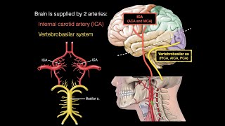
The Cerebral Arteries
The internal carotid artery
The middle cerebral artery
The anterior cerebral artery
The posterior cerebral artery
The vertebral arteries
The basilar artery
The Ventricles
The lateral ventricle
The anterior horn
The occipital horn
The temporal horn
The interventricular foramen (of Monroe)
The third ventricle
The cerebral aqueduct (of Sylvius)
The fourth ventricle
The lateral aperture (of Luschka)
The median aperture (of Magendie)
The checklist and quiz can be downloaded from the link below:
www.dropbox.com/s/jaekuxjwyma...
Please support this channel on Patreon: (website being verified)
www.patreon.com/user?u=793646...
DICOM Viewer is Horos from Horos Project:
horosproject.org
CT was made available for use for teaching at Embodi3D:
www.embodi3d.com
This exact MRI can be downloaded here:
www.dropbox.com/sh/hcu7im8zk3...
#Brain #MRI #anatomy


Amazing video. Thanks so much for your patience and love for teaching.
Great teaching style! Made it enjoyable and less boring
THANK YOU SIR YOU ARE A LIFE SAVIOR 🙏💗
Thank you for an amazing video!!
Great educational video.
Wonderful work I appreciate your effort
Thanks
Go on
May Allah bless you ❤
thaaaaaaanks very very much!!!! great job!! how unfortunate that so little people had watched and subscribed ur channel !!
u r a big help!! thanks very much, appreciate such a huge effort
Fascinating thank you
Was great, thank you !
Thanks
Any excellent vascular Types around? I've got a strange looking RVA that *looks like* it's kind of 'glued' to the side of the foramen magnum...on CT, 0.5 mm slice, the artery wall seems contiguous with the dura as it tranits the foramen magnum, just below a fairly large kind of ring-shaped calcification (reading 400-1000)...is this "attachment" to the cranium "normal"?
It was a great session...thank u very much
Thank you so much sir for making this video. I've looking for this type of video long time. Could you please tell me which application you are using in your mac?
Watching u from Syria
Big love❤
Thanks 💪🏽👍🏽♥️
You’re welcome 😊
Sir Please make a video of mri spine
Nice , ty
❤
Compression des ventricule lateral
Do you review brain mri sir ?
What mri is the 3t I'm guessing higher?
1:08:50 - Right Cerebral Peduncle
Correct. My fault. Not cerebellar peduncles, but ‘cerebral’ peduncles. Thanks for your input, truly appreciated.
Thnx sir, please what's this application that you use ?
I use Horos. It is an application for Mac only. It is free. The link is available in the description. I Have attached it below as well:
horosproject.org
Great presentation.... cerebral peduncle not cerebellar. at 1.07.28...thanks
La grandiose grande hypophise
The pituitary stalk appears hollow on imaging but in reality, the infundibular recess of the third ventricle extends in the Pituitary stalk, it's not hollow.
Thank you so much for this reply. I didn't know the answer to that question and I am very glad that you answered it. Appreciate it!
Thank you for the amazing video. @@alisradiologicalanatomycourse
The pleasure is all mine :) @@syedmahboob5045
On which imaging its hollow or hypointense ?
@@Dr-789 I should not have used the word "hollow". The infundibular recess is a part of third ventricle and should give CSF signals on respective imaging/sequence.
L'opacite dans le ventricule lateral?
Substance blanches et substance grise
Quesqu'il y'a dans le ventricule lateral?
Cn we have anatomy of brain on CT pls..
Brain tissue is not visualised on CT as well as it is on MRI. If one understands brain anatomy on MRI, it is very easy to translate that knowledge onto CT. I may not make a video of the anatomy of the brain on CT for this very reason. If there is any particular aspect of brain anatomy on CT you are interested in, let us know and I’ll try to address it. Thanks for your comment. It’s greatly appreciated.
Elli and elarab
Oui c'est chronic
Avec compression
Intradural
Extra dural est une urgence neurologique
Oedeme Aussi
Effet de masse
Malla artere basilaire
L'autre est une urgence
Qui control le corps par les hormones
Malla olfactif nerv
Mais oedeme tout autour
Chez les vieux
Ana ils m'ont restes que deux neurones fonctionelles
Kindly make video on dorsal stream visual pathway, I am confused on the issue of white fiber tracts which dorsal stream visual pathway employs
All nerve pathways involve white fibre tracts. The dorsal stream visual pathway is no different. Where it is exactly is a very specialised question which I will not be able to answer in a video. In short, it extends from the primary visual cortex in the occipital lobe to the posterior aspects of the parietal lobe. It is involved in spatial orientation and recognition.
Dear sir,
Kindly tell me one thing,,
Why initially Mri scan showed less white matter and later on huge tracts of white matter . Just like this Mri scan showed only few gyris initially, then huge number of gyris.
Hope you will reply my submission
I may have not understood your questions, feel free to rephrase it. Nonetheless, it is an important concept to understand the difference between gray and white matter. Gray matter is mostly un-myelinated interneurons while white matter is mostly myelinated neurons travelling larger distances in tracts. In the brain, in the peripheries, there is gray matter; and towards the centre, there is white matter. All the calculations, analysis and interpretation (perception, thinking and action) happens in the gray matter, while all the transfer of information to and from the brain happens in the white matter.