Unforgettable: Radiological Anatomy of the Hippocampus

Vložit
- čas přidán 12. 06. 2024
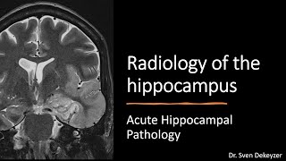
- The hippocampus is a small but complex anatomical structure that plays an important role in spatial and episodic memory. The hippocampus is located in the inner most medial part of the temporal lobe and is part of the limbic system. In this video I'll walk you through the anatomy of the hippocampus on MRI-studies. Knowlege of normal hippocampal anatomy is important to be able to detect hippocampal pathology, which will be adressed in two future videos. Want to know more? Check out this free access review article on anatomy and pathology of the hippocampus: insightsimaging.springeropen....
0:00 - Introduction
1:07 - Anatomy of the temporal lobe
4:50 - Macroscopic anatomy of the hippocampus
12:02 - Microscopic anatomy of the hippocampus
19:35 - White matter tracts of the hippocampus: alveus, fimbria and fornix
22:14 - Embryological development of the hippocampus: incomplete inversion
30:10 - Embryological development of the hippocampus: sulcal remnant cysts
31:12 - The function of the hippocampus: the Limbic System
33:29 - The function of the hippocampus: the story of patient H.M.
35:41 - Conclusion
This video is brought to you by "the Neuroradiolgist" :):
/ theneuroradguy
/ the_neuroradiologist
theneuroradiologist.org/
e-mail: neuroradiology.online@gmail.com
#neuroscience #neurology #radiology #meded #neuroradiology #neuroradiologist #theneuroradiologist #hippocampus #memory


Finally I found a clear description of the limbic system and hippocampus. Fantastic presentation.
really good presentation, thx a lot
This video helped me to fill many of my knowledge gaps regarding hippocampus. Thank you for sharing your knowledge. May God bless you, Thank you very much. Please continue the good work.
Extremely good 👍 😊 😮
Thank you!
Sir your lecture is most informative, clear and simple no one can replace you sir. Kindly do more neuro anatomy and pathology videos sir we will be waiting for more simplified and crystal clear videos sir.
Thank you sir
I’m happy to watch this video.
Great! I really enjoyed your presentation. These days, I get more and more interested in Hippocampus, especially from an evolutionary perspective.
Thank you very much for an amazing presentation. Highly appreciated.
Really made a difficult topic easy to understand, Requesting video on neuroimaging of Cranial nerves.
Clearly explained. Simplifies this complicated topic 😁
@thehneuradiologist... excellent, brillant and so so clear class!!! Thks. Federico (DrQ! - Psychogeriatrist & Neuropsychiatrist) from Buenos Aires, Argentina
Stratum lacunosum moleculare !!! 🤌😅
Thanks doc!!
Excellent explanation
Excellent simple and very clear presentation. I look forward to learning even more from the rest of your videos.
This is the most beautiful neuroanatomy video I have ever seen. You are wonderful & great teacher. Thank you so much for your effort. I would kindly ask you to be aware and lable left/right side hippocampus in the early images, as you have shown some times right and some times left hippocampus, which may lead to some confusion as how it appears regarding orientation of the cornu Ammonis. Pronunciation is not "hypocampus" with long i but hippocampus with short i).
Best regards
Thank you for your nice comments and interesting feedback, if I ever make a new recording of this presentation, I will definitely keep it in mind!
Wonderful, thank you very much for this clear explanation.
Wonderful explanation
I appreciate your effort
Thank you
Go on
May Allah bless you
AMAZING!!
Doc, wonderful video& a walk through hippocampus 👍Thanks for the great info.Can anything be spoken more on FORNIX?
Thanks for sharing your knowledge!
Great. Thankyou for sharing. God Bless
Amazing lecture❤ keep it up.
Wonderful lecture
Thank you sir
great lecture
thank you very much. Please continue doing these lecutres
useful lecture. Looking for part 2
Sven, super Vortrag, riesen Dank für deine Mühe. Warte gespannt auf Teil II. In guter Erinnerung aus den UKA Zeiten unter Prof. Wiesmann. Grüße aus Braunschweig
Thank you so much 🙏🏾❤️
Very nice,informative and clear
presentation.Thanks sir.Please give video lecture on Amygdala.
great lecture ! very informative
Thanks!
excellent merci
🙏 very useful
and when the hippocampus works at 100% efficiency, you remember who you are... hence, Amon
Pięknie!
Sir, kindly make videos on MRI spine radiological anatomy
❤❤❤❤
Please provide timestamps
Done, thanks for pointing out they were missing
Hello 😉
excellent lecture. Only issue is the nomenclature for the lateral temporal sulci. Most authors would list the temporal sulci as 1) superior temporal sulcus, 2) inferior temporal sulcus, 3) occipital-temporal sulcus and 4) collateral sulcus.
Thank you for you comment! Could be. Something I noticed is that with anatomical nomenclature different authors and handbooks sometimes use different names for the same structures (e.g. fusiform gyrus vs. lateral temporo-occipital gyrus). Could you give me a reference to the handbooks or authors you are using? The handbooks and authors I follow all describe three lateral sulci (superior, middle and inferior), and I would like to compare sources.
@@theneuroradiologist I cannot provide a standard reference. Rather a representative example of how the temporal sulci are named in the fMRI community
www.ncbi.nlm.nih.gov/pmc/articles/PMC3138128/
In this paper the authors state that the human homologue of the MT region is located along the ascending limb of the posterior part of the inferior temporal sulcus, which is very standard language in the field. If you look at the images it is clear that they refer to the sulcus along the upper border of the inferior temporal gyrus.