Meninges and haematoma (anatomy)

Vložit
- čas přidán 20. 07. 2024
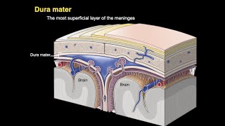
- We're going to look at the anatomy of the meninges covering the brain and describe the pia mater, arachnoid mater, and dura mater. We're also going to look at where the blood vessels inside the skull run relative to these layers so that we can confidently talk about extradural (or epidural) haematomas, subdural haematomas, and subarachnoid haemhorrhages.
For more about the dura mater: • Dura mater
For more about dural venous sinuses: • Dural venous sinuses
For more about the ventricles of the brain and cerebrospinal fluid: • Ventricular system of ...
For more about the blood supply to the brain: • Blood supply to the br...
For more about the middle meningeal artery and extradural haematoma: • Middle meningeal arter...
Music by Jahzzar
Album: HiFi City Tales
Song: Bodies
jahzzar.bandcamp.com - Věda a technologie


Keep recording such amazing lectures👍
I had a oral Anatomy exam last week and you helped me so much! Thank you Sam
Greetings from Iceland
Been using your lectures as learning material on my drives to and from medical school in the US. Lived in the UK for the first 16 years of my life and you take me back to the days of school in England. Thanks so much for what you do sir! Please don’t stop
One of the best lectures I have seen on anatomy. This guy is also so passionate about the human body and it truly shows. Thank you for the amazing content. Pre-med student here.
❤
Thanks God 🙏 u. R back keep going Dr Sam. U r the Ray of hope fr us
It's a long time since I last heard someone use the word bleb (blister) ...shows I was paying attention tho! I feel this is the calm before the storm...let's see how long it is before frenetic whirly tutor is back. Respect!
Thank you so much for these lectures Dr.Sam, it really is hard to find quality knowledge and such precise yet easily comprehensive teaching.
Your lectures really are extremely valuable to thousands of medical students around the world.
thank you for this lesson! i lost it when you said "spider mother". i finally got the deal about the arachnoid granulations.
Outstanding way of teaching Sir... we wanna to thanks of you Sir
Sir you make Neuroanatomy way easier ! Our seniors say its the most difficult part of human anatomy but you make it so much interesting that I actually want to learn more !!
You are a life saver for me... This not craming, but learning the real thing.
Fascinating, thankyou. I keep growing meningiomas. My first one was the size of a large lemon and was blocking half of my spinal fluid with my brain pushing down into my neck so the surgeon was keen to remove it asap. I had a left occipital craniectomy in 1998. I have a large part of my skull missing there. It was after that operation I had massive headaches for many years. I then started to grow a 2nd one, small, it was gamma knifed in 2007. The 3rd one small again I had 28 day proton therapy. Those 3 procedures were done in the US when I lived there. I moved back to Australia and in 2017 an MRI showed I was growing 2 more. I can't have gamma knife as they say you can't gamma knife the same area twice due to the splay of radiation. Proton is the least invasive but we don't have that here yet. They hope to have one built in 2 more years. A lot of info, however I'd love to hear your information on meningiomas if you are interested in sharing a video about them.
Good luck. I did think about including meningiomas in this video but thought it would be a little too much for the topic. I'm not sure I know enough to make a whole video about meningioma.
@@SamWebster Thanks Sam, any info you gather I'd appreciate watching. I'm a visual person so it's really helpful. I've enjoyed the couple of videos I've seen.
I love how passionate you are about what you teach, Thank you sir :)
How humble you are.
You probably won‘t see this, but… Dura mater does have two layers around the spinal chord (stratum periostale and meningeale), they are just separated and there is a space between them filled with fat, I believe some vessels and connective tissue.
Sumatriptan works decently for me. I suffer from migraines 3 to 4 a week. Just started trying Nurtec, after my 2nd dose I felt migraine free for 5 days as if I never had them, then the migraine pain came back like out of nowhere. It also makes me sleepy and have nightmares. Thanks for your professional wisdom and insight. I wish there was a machine like a blood pressure machine that could actually tell you just how much pain you are in because sometimes it's indescribable off the charts, ice picking, shattered glass on your brain kind of pain. Also just had my second dose of Botox 32 shots total, may work over time... I shall see, chronic pain and chronic illness isn't worth living with over time.
Outstanding sir.... easy way of teaching... the best one
you helped a lot in my head and neck anatomy course !
😊Your vedios are undoubtedly amazing full of knowledge..thank u so much sir ur vedios are very helpful🙌lots of love from India❤😇
Thank you for a wonderful lecture. Very well explained 👍
So much awaited topic
It is needed sir thank you for uploading this 🙏🏼🙏🏼🙏🏼🙏🏼🤩🤩🤩🤩
You are the best teacher ever!!
Great in depth explanations!!
I am really into this way of explaining anatomy. Cool!
This is one of your best videos! Excellent
Fantastic job as usual!
Your videos are so beneficial, plz keep making such videos
Awesome as usual , thank you so much 🙏
Thanks mate/doc !
Very interesting indeed plus an excellent explanation 👍
Cheers
Really wow! You are amazing! I watched a couple of actual brain dissection and neuro-surgery videos, and with those videos and yours, I learned a lot. The best thing is that you made me smile and laugh sometimes, making listening easier. Thank you.
thank you for such amazing videos! its really helpful as i am a med student, first year, at home, without any dissection trying to do head neck brain anatomy!! its soooo difficult.. and you make it so much easier! and not to mention, you look like house m.d [hugh laurie] and you have almost the same expressions and verbal skills as him! okay coming to the more important part.. pls do some embryology videos too! we often overlook embryology for our exams and even in classes thinking it carries very little weightage, but even a little weightage is a lot in this field! embryology videos would realllllllyyyy help. thanks agaiiin!
There are some embryology podcasts here: samwebster.net/?page_id=2538. I'm not planning on making any embryology videos in the near future. This textbook is alright too: www.amazon.com/Embryology-at-Glance-Samuel-Webster/dp/111891080X
@@SamWebster thank you! It's a great help. Embryology is always difficult. Dont you think? What about histology.?
@@divyavachhani6963 I try to include histology in my videos where I can. The microscopic and macroscopic all make up the same structure and contribute to its function.
@@SamWebster thank you for your prompt reply! I appreciate your efforts to educate all of us!! I passed my midterms with your videos! Things that felt so difficult.. I could just easily visualize without even memorizing.. and it was amazing!!! Thank you thank you thank you!
Thank you for another amazing video! I have a question, where does the fluid go after leaving the skull? What happens to it after it is back in the systemic circulation?
Does anyone think he looks like HOUSE MD(Hugh Laurie)😂♥️
I was looking for this comment 😅💜💜
omg one of the reasons, i stumbled across him!
thank you these videos are amazing!
Thank you so much for these videos - I passed my musculoskeletal units in medical school during the pandemic ENTIRELY because of your videos. Do you have any recommendations for additional resources we can use now that we can't attend anatomy wet labs/use models at our university?
All I do is talk - you have to think and remember, so I'm sure your results are because of the work you put in. I would recommend one of the 3D CGI anatomy apps like Visible Body or 3D4Medical for more study. You can get a better idea of how structures lie in relation to one another than from a textbook alone.
Hi, your videos are brillian. One question, you said in the model that we can see the veins so it must be the arachnoid. I'm confused, so one can seperate arachnoid from dura?
Thanks for this sir...Neuro anatomy is an issue for me!😅
I've never understand what happens to the Meninges at the Cauda Equina. I know the meninges end there, but what exactly does this look like?
It pulls to a point at the inferior end of the spinal cord and continues as a strand (the filum terminale) to the coccyx.
Cool....keep posting...u r awesome 🤝
God bless you doc...👆
Thank u so much ❤️
thank you so much your the best
Super!!!! 👍 God Bless
Very beautiful .....
Thanks Sam! :)
Don't you just hate it when he says "There's more to it than that..." Makes you think "What have I missed???" 😂
아주 좋아! 1등이닷!
Sir retinaculum of lower limbs especially ankle joint
Which structure will pass from
Getting confusion
How does a headache occurre with the subarachnoid haemorrhage, since the dura mater is the one sensitive to pain? I assume it has something to do with the change in pressure. I would be very appreciative if someone could answer :)
Youre a godaamn herrooo!!!
💙💙
You remind me of dr. House
memorising stuff from a lecture powerpoint takes time but truely understanding something is another thing which will help me memorise my stuff. Thank you.
This guy is volunteer
I luv u
Anybody told you before you look EXACTLY like Gregory House😯
Once or twice. I broke my foot and had a stick to walk with once, which drew even more comparisons.