Femoral Nerve Anatomy Animation : Origin, Course, Sensory and Motor innervation, Clinical anatomy

Vložit
- čas přidán 10. 05. 2024
- 📌 𝐅𝐨𝐥𝐥𝐨𝐰 𝐨𝐧 𝐈𝐧𝐬𝐭𝐚𝐠𝐫𝐚𝐦:- / drgbhanuprakash
📌𝗝𝗼𝗶𝗻 𝗢𝘂𝗿 𝗧𝗲𝗹𝗲𝗴𝗿𝗮𝗺 𝗖𝗵𝗮𝗻𝗻𝗲𝗹 𝗛𝗲𝗿𝗲:- t.me/bhanuprakashdr
📌𝗦𝘂𝗯𝘀𝗰𝗿𝗶𝗯𝗲 𝗧𝗼 𝗠𝘆 𝗠𝗮𝗶𝗹𝗶𝗻𝗴 𝗟𝗶𝘀𝘁:- linktr.ee/DrGBhanuprakash
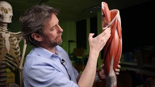
Femoral Nerve Anatomy Animation
The femoral nerve, a major nerve arising from the lumbar plexus, originates from the dorsal divisions of the ventral rami of the second, third, and fourth lumbar nerves (L2, L3, L4). It emerges through the psoas major muscle, descending between the psoas and iliacus muscles, and then travels beneath the inguinal ligament into the anterior thigh. This nerve is crucial for the innervation of the quadriceps femoris muscle group, which is key to knee extension. Additionally, it provides sensory innervation to the anterior thigh and medial aspect of the lower leg via the saphenous branch. Given its vital role in leg movements and sensation, the femoral nerve's integrity is essential for walking and maintaining balance.
#fmge #fmgevideos #rapidrevisionfmge #fmgejan2023 #mbbslectures #nationalexitexam #nationalexittest #neetpg #usmlepreparation #usmlestep1 #fmge #usmle #drgbhanuprakash #medicalstudents #medicalstudent #medicalcollege #neetpg2023 #usmleprep #usmlevideos #usmlestep1videos #medicalstudents #neetpgvideos #FemoralNerve #Anatomy #LumbarPlexus #Neurology #MedicalEducation #MedStudent #Physiology #HealthEducation #Biomechanics #Orthopedics #SportsMedicine #PhysicalTherapy #MedSchool #MedicalScience #HumanAnatomy #anatomyandphysiology


Thanks from somalia
Good lesson. Thanks.👍
Glad you liked it!
Can u plz do for liver
Nice video
So nice
M,.