Brain imaging course - 1 - Imaging Modalities

Vložit
- čas přidán 19. 05. 2024
- This video is the first in a series of a brain imaging capstone course to learn some of the basics about brain imaging. The overall series will cover the range of imaging used to investigate the brain, information about how to choose what type of study will help your patient, teach you how to review images on your own, review some common pathology, and then provide some interactive courses that you can review on your own.
The entire course is available here:
learnneuroradiology.com/brain...
0:00 Introduction
2:05 Modalities used
The main types of imaging, or modalities, used in brain imaging, are computed tomography (CT) and magnetic resonance imaging (MRI). Each of these can be tailored in specific ways to look at vessels, including arteries or veins.
2:46 CT head without contrast
CT head is the main screening exam used in neuroradiology. This is commonly done any time a patient has new neurologic symptoms and can see common pathologies such as stroke, hemorrhage, fracture, edema, and hydrocephalus. Once patients are in the hospital, it may be used to follow up their pathology.
3:23 CT head with contrast
While possible, we almost never perform CT of the head with contrast because MRI is a much better examination and will almost always be done anyway.
4:24 CT angiogram
CT angiogram, or CTA, is an arterial timed exam to look at the arteries of the brain. This is very commonly done in evaluation of stroke, intracranial hemorrhage, and trauma. Aneurysm and vascular malformations are very well evaluated by CTA.
4:57 CT venogram
A CT venogram, or CTV, is very similar to a CTA, but the timing is a little later. This is optimal for evaluating the veins of the brain for thrombosis or trauma.
5:20 X-rays
We don’t use many x-rays in neuroradiology, but you may see a few to evaluate for shunts and hardware. CT is almost always better, particularly in trauma.
5:58 MRI brain
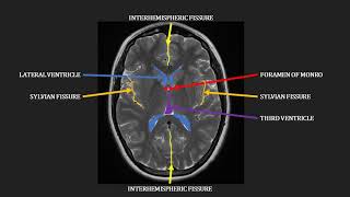
MRI of the brain is a workhorse of neuroradiology. It has great tissue contrast and is excellent for finding diseases of the brain. Some limitations include availability/expense, limitations in patients who have devices, and the time that it takes. There are a variety of sequences that we use in MRI of the brain, and each tells us a little bit of something different about the brain.
7:30 T1 precontrast
The T1 precontrast images are useful for evaluating the overall brain structure and alignment. It is also useful for comparing to postcontrast imaging to see how much enhancement there may be.
8:01 T2/FLAIR
T2 images are water-sensitive images on which most pathology will show up as bright. It is great for looking at edema, swelling, and fluid-filled structures. FLAIR images are very similar to T2, but the fluid has been suppressed. This helps pathology be more obvious and easier to detect.
10:04 Diffusion (DWI)
This is a measure of how well water moves through tissue. In stroke, water moves into cells and can’t move as freely, resulting in areas of stroke being bright on DWI.
10:34 Blood sensitive imaging
Gradient imaging (GRE) or susceptibility weighted imaging (SWI) provide a chance to better detect calcium and blood, which will appear dark.
11:07 T1 postcontrast
These T1 images are obtained after an intravenous contrast agent has been administered. Things that enhance, or are bright on these images but not the precontrast images, accumulate contrast. This often occurs in pathologies like tumors because the blood-brain barrier has become leaky.
12:06 MRA head
MRA of the head is (most frequently) a noncontrast technique to evaluate the vessels of the brain. This is a great technique to see the vessels of the brain if you are not in a rush, particularly to see aneurysms and vascular malformations.
12:38 MRA neck
Similar to MRA of the head, this is vessel imaging of the neck. You can do it without contrast or with contrast, but contrast often helps see the vessels at the thoracic outlet better.
13:07 MR venogram
Like a CT venogram, an MR venogram is a dedicated exam to look at veins to look for venous thrombosis or venous injury.
13:30 Summary
Thanks for tuning in to the video. Hopefully you learned a lot about the types of imaging used to evaluate the brain.
Be sure to check back in for additional videos in the future or check out the website at
www.learnneuroradiology.com


Excellent video. Looking forward to the whole series.
Awesome, thank you!
Excited to attend the future lectures sir! Thank you for this!
Thank you for your great lectures.