Imaging Flow Cytometry: A Brief Overview - Andrew Filby (Newcastle U.)

Vložit
- čas přidán 21. 04. 2019
- www.ibiology.org/techniques/i...
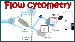
In this talk, Dr. Andrew Filby provides an overview of imaging flow cytometry, a powerful technique used to measure the phenotype of cells using image-based metrics. Compared with traditional flow cytometry, imaging flow cytometry increases the number of parameters one can measure by providing morphological and spatial information in a high-throughput, controlled manner. Filby explains how the imaging flow cytometer works and describes the benefits of using this technique to better answer biological questions, as well as giving us a glimpse into the future of this exciting field.
Speaker Biography:
Dr. Andrew Filby is the Director of the flow cytometry core facility at Newcastle University in the UK. He received his bachelor’s degree in biochemistry from the University of Huddersfield, and his Ph.D. in molecular and cellular immunology from the National Institute for Medical Research (NIMR) in Mill Hill, London. Filby joined the laboratory of Dr. George Kassiotis at the NIMR where he continued his post-doctoral training. After his post doc, he joined the London Research Institute as the deputy head of the cytometry core facility. Filby continues to innovate and develop new cytometric applications in his current position at Newcastle University. - Věda a technologie


![[柴犬ASMR]曼玉Manyu&小白Bai 毛发护理Spa asmr](http://i.ytimg.com/vi/0TsXQ7z2Dh4/mqdefault.jpg)


Clear, concise and informative: an excellent video.
This is fantastic, should be way more popular
Great presentation and very clear explanations. Thank you
Very detailed and well explained thank you
I learned a lot from this, Thank you!
Great presentation ! thanks
Thanks a lot for the efficient explanation
Hello, this video is very clear but I hace a question. Are we seeing the memebrane af the cell or a slice of it?
Just like in microscopy, it has a focus, and using a focus you see a "slice of the cell" just like how a focus on a normal digital camera allows you to see different depths.
Fantastic video
Adding images to flow cytometry. (12:12,25:00)
Nice presentation sir......next time ..plz deduce ur speed of presenting 👍
WAW! What a presenter...
What is the difference between 'imaging flow cytometry' and 'full spectrum flow cytometry'? If there is any? 😬
bravo