Femoral Triangle - Everything You Need To Know - Dr. Nabil Ebraheim

Vložit
- čas přidán 26. 02. 2015
- Dr. Ebraheim’s educational animated video describes the Femoral Triangle of the Thigh.
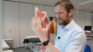
The femoral triangle is a superficial triangular space located on the anterior aspect of the thigh just inferior to the inguinal ligament.
The boundaries of the femoral triangle include:
1-Lateral border: formed by the medial border of the Sartorius muscle.
2-Medial border: formed by the medial border of the adductor longus muscles.
3-Base: formed by the inguinal ligament.
The floor of the triangle is formed by:
1-Iliacus muscle
2-Psoas major muscle
3-Pectineus muscle
4-Adductor longus muscle
The roof of the femoral triangle is covered by skin, superficial fascia and deep fascia.
Content: the femoral triangle contains three important structures: from lateral to medial:
1-Femoral nerve
2-Femoral artery
3-Femoral vein
4-And it contains deep inguinal lymph nodes.
Navigate the femoral triangle from lateral to medial.
The femoral triangle also contains the femoral sheath which is a funnel shaped sleeve of fascia enclosing the upper 4 cm of the femoral vessels. The femoral nerve is the most important nerve within the triangle. However, it is not the only nerve. The femoral nerve lies within the groove between the iliacus and the psoas major muscles.
Two other nerves are located within the femoral triangle:
1-Lateral cutaneous nerve of the thigh: crosses the lateral corner of the triangle and supplies the skin on the lateral part of the thigh.
2-Femoral branch of the genitofemoral nerve: runs in the lateral compartment of the femoral sheath and supplies the majority of the skin over the femoral triangle.
The neurovascular bundle is media to the Sartorius muscle. Therefore, in the anterior approach to the hip, it is always safe to go lateral to the Sartorius muscle to avoid the important structures within the femoral triangle. It is important to remember when performing this approach to avoid the lateral cutaneous nerve of the thigh.
The femoral sheath is a fascial sheath that contains the femoral artery, vein and femoral canal. The anterior portion of the femoral sheath is formed by a downward extension of the fascia transversalis. The posterior portion is formed by the iliac fascia. The femoral sheath is divided into three compartments. The lateral compartment contains the femoral artery. The intermediate compartment contains the femoral vein. The medial compartment is called the femoral canal and contains lymphatic tissue.
The base of the femoral canal is formed by the femoral ring. A femoral hernia occurs when part of the intestine protrudes through a weak femoral ring into the femoral canal.
Become a friend on facebook:
/ drebraheim
Follow me on twitter:
#!/DrEbraheim_UTMC
Donate to the University of Toledo Foundation Department of Orthopaedic Surgery Endowed Chair Fund:
www.utfoundation.org/foundati...


Thank for your clear description.
Got exactly what i needed thank you very much sir
Thank you!!!
Thanks ur vedios are helping me very well
I apreciate for providing such a good videos
Thanks you very much sir ,your videos are easy to understand
Loved it, much concise and helpful💯
Amazing......So easy to understand........Thank you....
super, thank you...
thank you very helpful for me , while I'm studying about femoral hernia
THANKU SO MUCH FOR MAKING SUCH VIDEOS FOR US, IT REALLY MAKES EVERYTHING EASY, I ALWAYS PREFER YOUR VIDEOS. :)
thank you!
Thank You very much
What a great video!💖💖💖💖💖💖💖
thank you soo much very easy and nice video...
amazing
You have a stunning sound . And the content is really good too. Subscribed.
So Thanx^^
excellent video from brilliant person , you saved my time searching for every part on google to see its location
Thanks Doctor you are awesome ! :)
Excellent explanation with a good quality video..💖
Really good for understanding sir 👌👍, Thank you sir
osmmmmmmm than osmmm it's actually .....i loved it ...n it's very helpful to me thanks alot
Actualy i got what i need..in this videos...very nice sir.
Thankyou...very much informative
This makes studing so much easier!! Brilliant!!
thank you very very much 😢
وشك شبه الطشت
Gracias . Tus vedios ahelGracias . Tus vedios me interesan mucho. Deseos de India
merci bien
Best💖
What is the medicines this femoral triangle
doctor could you please help me ?
about the medial border of the femoral triangle ; why is it formed by the medial border of the adductor longus muscle instead of the lateral border of the aductor longus muscle ?
same question here
please make the next video with ur voice such that
Superlike
One of the few videos detailing the ring. Thank you. I am curious is the four cm the sheath that bind all 3? Terrific job. Thank you.
and now you made me question it too
okay now that I’ve searched… (although I don’t fully understand that question) the femoral sheath contains the femoral artery, vein and deep inguinal lymph nodes and vessels and each one of these is separated by fascial compartments within the sheath so yes it does wrap these 3 together along its 4cm
@@trulyasleep294 I’m trying to understand it in terms of hernias in particular.
Thigh Triangle!
NAVEL - Nerve Artery Vein Empty Space with Lymph Nodes
tank's
Those that appendix edc say hi.