Anatomy Of The Popliteal Fossa - Everything You Need To Know - Dr. Nabil Ebraheim

Vložit
- čas přidán 30. 07. 2018
- Dr. Ebraheim’s educational animated video describes the anatomy associated with the popliteal fossa - posterior knee.
Follow me on twitter:
#!/DrEbraheim_UTMC
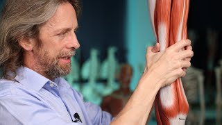
The area of depression located at the back of the knee joint is called the popliteal fossa. The popliteal fossa is a closely-packed space. It is bounded by the biceps femoris laterally, as well as the semitendinosus and semimembranosus medially. The lower part of the space is formed by the two heads of the gastrocnemius muscle.
Four common conditions involving the popliteal fossa include:
1. Baker’s Cyst
2. Popliteal Artery Entrapment Syndrome
3. Posterior Knee Dislocation
4. Posterior Cruciate Ligament (PCL) Injury
A Baker’s cyst is a benign swelling found behind the knee that lies between the semimembranosus and the medial gastrocnemius muscles. A Baker’s cyst is also known as a popliteal cyst which lies posterior to the medial femoral condyle. The cyst is connected to the knee joint through a valvular opening. Knee effusion from intra-articular pathology allows the fluid to go through the valve to the cyst in one direction.
Popliteal artery entrapment syndrome is a rare condition involving extrinsic compression of the popliteal artery behind the knee due to the anomalous relationship of the muscle and artery in the popliteal fossa. It may also be caused by fibrous tissue constricting the artery. This condition usually affects younger athletes who present with calf claudication. The blood flow will be decreased. The patient will complain of swelling, foot numbness and paresthesia, tingling of the toes, and cramping of the muscles. Plantar flexion of the ankle and hyperextension of the knee will decrease the pulses. An arteriogram is probably the best study showing the compression and condition of the artery. Treatment consists of observation and activity modification. Surgery may be necessary to release the muscle and relieve the pressure on the artery.
Posterior knee dislocation occurs as a result of violent trauma. The most common mechanism of injury includes exaggerated hyperextension of the knee and dashboard injuries. Posterior knee dislocation may be associated with a high incidence of popliteal artery injury.
Posterior translation of the tibia will occur with rupture of the posterior cruciate ligament. A common cause of this injury is a bent knee hitting a dashboard during a car accident; however, it occurs more frequently in sports from forced hyperflexion of the knee.
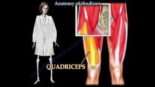
The quadriceps active test is conducted to test the quadriceps muscle. The examiner stabilizes the leg of the patient and then the patient is asked to actively contract the quadriceps muscle. The tibia is seen being actively reduced from the posterior subluxed position. The Lachman’s Test is another test used to examine the knee. When performing the Lachman’s test, the knee is bent at 20-30°. The examiner provides posterior force to the tibia while applying anterior pressure to the femur in order to access the posterior translation of the tibia. The posterior drawer test is carried out while the patient is in a supine position and the knee is flexed to 90°. The amount of translation of the tibia relative to the femur is observed. The dial test is performed while the patient is in the supine or prone position and both knee are in 90° (it shows the PCL injury) and 30° of flexion (will show the posterolateral corner injury). More than 10 degrees of external rotation indicates significant injury.


We should also add that the contents of popliteal fossa also includes the Geniculate branch of the obturator nerve as well the Posterior cutaneous nerve of the thigh🦵🏻
Ur videos are very much helpful i covered many of my topics from ur videos thanks and keep posting 👍😊
Thank you , it is great to know such information
Very useful information!! 🧠 Thanks doc!! 🙃
I want to give u one million likes thank u so much sir
Thanks for this amazing videos plz keep posting
Very helpful ...Thank you sir
Thank you
Thanks a million stay happy
Thank you very much
Thank you very much ,sir..
Thank you proff Ebraheim
Thank you it was very helpful
Thanks, helpful !
Very helpful
Thank you sir
thankyou so much
Thanks a lot!
Thanks Dr
thanks a lot
Thanks sir😊🌱🙏
Niceone Mr.
Thank u sir
Thanks for this video.May Allah bless you Sir.
Thanks sir
Key samg aaya
ครับผม ด้วยความเคารพ รักษาสุขภาพเข้มแข็ง มีความสุข ครับผม คุณครูที่รักษ์ สาธุการณ์ครับผม ด้วยความเคารพ ครับผม
Thanx too much doc
Tq sir
Yes, thank you, but what about the pain? It hurts when my knee is bent back towards my torso and lifting up. It's my left left knee and when I put my left leg in the drivers door, the knee is weak and painful. I use my hand to lift that leg into the car. I had this pain in the right knee a couple of years ago and the pain disappeared after about a year of pain. I am a walker or used to be. I don't walk 10k's hardly at all now. Thank you.
Good
This is what im talking about. Do we have a deal!!?
❤️❤️❤️
Jklq Dr
Tq sir