Cranial Bones, Sutures and Bony Landmarks | Skull Bone Anatomy | Radiology Anatomy Part 1 | CT Brain

Vložit
- čas přidán 17. 06. 2024
- Let's revise the anatomy of the neurocranium. Get the companion document here: bit.ly/anatomydocument
Advanced mock past paper anatomy exams using clinical images 👉 www.radiologytuts.com/courses...
Thank you to SCP radiology for the images provided
/ scp-radiology
scp_radiolo...
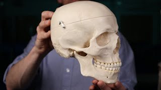
Want to check out the anatomy models used in this video?
Cervical spine: hubs.li/Q02mB_s_0
Skull model: hubs.li/Q02mB_Pz0
Anatomy Warehouse: hubs.li/Q02mC02W0
Use the coupon code: RT_15Off_Axis for 15% off (this is not an affiliate link)
Studying for a radiology physics exam?
Get high yield radiology physics past paper questions with video answers
Perfect for testing yourself prior to your radiology physics exam 👇
➡️ X-RAY AND ULTRASOUND BUNDLE (SAVE over 25%): www.radiologytuts.com/bundles...
➡️ X-RAY QUESTION BANK: www.radiologytuts.com/courses...
➡️ ULTRASOUND QUESTION BANK: www.radiologytuts.com/courses...
➡️ MRI QUESTION BANK: www.radiologytuts.com/courses... 🕰️
=========================
I have also created three RADIOPAEDIA LEARNING PATHWAYS
bit.ly/radiopaediaphysics (👈 25% OFF DISCOUNT LINK)
WHAT’S INCLUDED?
✅This CZcams series Ad free
✅Constantly updated Radiopaedia articles
✅Summary slides
✅Key take home bullet points throughout
✅Multiple review quizzes
✅Short answer review questions
✅Official Radiopaedia course completion certificate
25% discount using this link: bit.ly/radiopaediaphysics
=========================
Join the Radiology Tutorials Newsletter. Short, simple, informative. 👉 bit.ly/3ruLh3d
=========================
Video chapters:
00:00 - Introduction
00:24 - Frontal bone
00:47 - Coronal suture
00:57 - Sagittal suture
01:02 - Bregma
01:23 - Persistent metopic suture (not visualised)
01:52 - Sagittal sulcus
02:07 - Frontal crest
02:24 - Frontal sinuses
02:50 - Superciliary arches
03:04 - Glabella
03:42 - Nasal bones
03:49 - Frontal sutures
04:06 - Nasion
04:22 - Supraorbital plate
04:43 - Supraorbital margin
04:37 - Supraorbital notch/ supraorbital foramen
04:55 - Supratrochlear notch
06:06 - Zygomatic process of the frontal bone
06:18 - Frontozygomatic suture
06:52 - Ethmoid bone
07:00 - Cranial fossae
07:20 - Ethmoid sinuses
07:33 - Crista Galli
08:01 - Olfactory fossa
08:16 - Cribriform plate
08:40 - Vertical lamella
08:47 - Fovea ethmoidalis
09:22 - Sphenoethmoidal suture
09:26 - Sphenoid bone
09:49 - Parts of the sphenoid bone
10:03 - Sphenoid body
10:24 - Sphenoidal yoke (planum sphenoidale)
10:29 - Sella turcica
10:35 - Tuberculum sella
10:42 - Hypophyseal fossa
10:48 - Dorsum sella
11:07 - Posterior clinoid processes
11:13 - Carotid sulcus
11:33 - Lesser wings of the sphenoid
11:54 - Superior orbital fissure
12:22 - Anterior clinoid processes
12:36 - Optic canal
12:55 - Optic chiasm
12:58 - Chiasmatic sulcus
13:16 - Greater wings of the sphenoid
14:43 - Foramen rotundum
15:31 - Pterygopalatine fossa
16:04 - Foramen ovale
16:30 - Foramen spinosum
17:21 - Sphenofrontal suture
17:37 - Spheno-occipital synchondrosis
17:46 - Sphenoparietal suture
17:49 - Sphenosquamousal suture
17:58 - Temporal bone
18:15 - Companion document linked below
18:27 - Temporal bone parts
18:41 - Squamous part of temporal bone
19:02 - Zygomatic process of the temporal bone
19:21 - Squamous suture
19:30 - Petrous part of the temporal bone
19:39 - Carotid canal
19:55 - Foramen lacerum
20:05 - Superior petrosal sinus
20:18 - Arcuate eminence
20:25 - Internal acoustic meatus (auditory canal)
20:34 - Sigmoid sinus
20:47 - Jugular foramen
21:03 - Mastoid process
22:35 - Parietal bone
23:10 - Lambdoid suture
23:22 - Lambda
23:51 - Pterion
24:22 - Asterion
24:33 - Occipital bone
24:42 - Occipital bone parts
24:59 - Cruciform eminences
25:07 - External occipital protuberance
25:23 - Foramen magnum
25:37 - Clivus
25:55 - Basion
26:00 - Opisthion
26:28 - Occipital condyles
26:49 - Atlanto-occipital joint
26:56 - Hypoglossal canal
27:12 - Jugular tubercle
27:42 - Inferior petrosal sinus
28:00 - Test yourself with real cases linked below


Man!!!!
You are very eloquent and very good in all your tutorials but by inckuding anatomy models you have REALLY taken your tutorials to a whole new level and have outdone yourself.
I wonder if you could include the anatomy models with moving grid lines back and forth like you do on CT scan coronal axial sagital views!!! That would make it all EVEN A GREATER!!!!
Thanks a lot for these anatomy models.
This is one of the best videos I've ever seen on this topic. Radiology trainees everywhere greatly appreciate your hard work Michael!
The best radiology resource in CZcams. Thank you for the effort.
Thank you! So glad the videos are helpful 😊
Sir Michael we are very excited for this new series 🎉
Hope you enjoy it!
youtube.com/@RadiologyinfoDailycases-ze?si=XZWmF_P4tl7kLjuH
Please never stop uploading 🙏🥺
Hi, I watched your MRI physics course on radiopaedia and I just wanted to thank you for all the effort you put into it. I've watched a lot of videos on MRI and your videos are by far the best. Finally it all made sense to me!
Wow, thank you. This means so much to me. Appreciate all your support
THIS WAS AMAZING. You’re the MOST BRILLIANT radiology educator out there. Thank you for making such videos..
Ah, thank you 😊
youtube.com/@RadiologyinfoDailycases-ze?si=XZWmF_P4tl7kLjuH
Honestly have gotten more out of your channel that most other anatomy sources I've tried to use in rads training - thank you for continuing this great work!
youtube.com/@RadiologyinfoDailycases-ze?si=XZWmF_P4tl7kLjuH
Super helpful thanks for sharing!
youtube.com/@RadiologyinfoDailycases-ze?si=XZWmF_P4tl7kLjuH
You are SO SMART!
Thank you so much for sharing. Radiology anatomy series are so helpful to me. 💯❤️
I'm so glad they're helpful! More to come soon 😊
very comprehensive. Thank you very much
youtube.com/@RadiologyinfoDailycases-ze?si=XZWmF_P4tl7kLjuH
Commendable job 🎉
Thank you indeed for fantastic educative presentations. I really appreciate your work and effort. Please continue and let it to be free.
Thank you so much for the hard work you put into making these videos! You have the best radiology videos on youtube!
Can't wait for the full series 🔥🔥
I can see the effort, the quality of the videos, thank you very much!
youtube.com/@RadiologyinfoDailycases-ze?si=XZWmF_P4tl7kLjuH
Superb lecture 👌 👏 👍 🙌
youtube.com/@RadiologyinfoDailycases-ze?si=XZWmF_P4tl7kLjuH
Thank you
Thanks for this series…. Kindly teach us ultrasound anatomy as well
It's really impressive sir❤
Thank you!
God bless you, handsome man.
will like and subscribe to everything you make so hopefully it can become financially rewarding enough for you to make more and more. thank you
You are amazing. Gifted and may the Good Lord bless your talent
Awesome
Hello , execellent teaching , the document isn't available !
Thank you ❤ please can u do a DSA tutorial
youtube.com/@RadiologyinfoDailycases-ze?si=XZWmF_P4tl7kLjuH
Sir mri lecture completed or coming more
Sir Michael we are desperately waiting for the next video of this series
youtube.com/@RadiologyinfoDailycases-ze?si=XZWmF_P4tl7kLjuH
Hii sir..can I have a chat with you because I have some doubts on mri..
I have university exam next week... please help me sir..😢
Please sir...