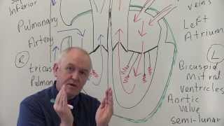
Introduction to Cardiovascular System - General anatomy

Vložit
- čas přidán 22. 08. 2024
- 📌𝐅𝐨𝐥𝐥𝐨𝐰 𝐨𝐧 𝐈𝐧𝐬𝐭𝐚𝐠𝐫𝐚𝐦 :- / drgbhanuprakash
Introduction to Cardiovascular System
The circulatory system, also called the cardiovascular system, is a vital organ system that delivers essential substances to all cells for basic functions to occur. The system supplies nutrients to and removes waste products from various tissues of the body.
The cardiovascular system is a network composed of the heart as a centralized pump, blood vessels that distribute blood throughout the body, blood serves as a medium of transportation, and the blood of different substances.
Blood vessels
-----------------------
The blood vessels form a closed system of tubes that carry blood away from the heart to the tissues of the body and then return it back to the heart.
The blood vessels include: Arteries. Capillaries, Veins
Arteries (distributing channels) are thick-walled tubes that carry blood away from the heart. The blood leaving the heart passes through the vessels of progressively smaller diameters referred to as arteries and arterioles
Capillaries are microscopic vessels that connect arterioles and venules.
Veins (draining channels) are thin-walled tubes that carry blood from tissues of different parts of the body back to the heart. The blood returning to the heart from the capillaries passes through the vessels of progressively larger diameters, termed as veins and venules.
Except for the capillaries and the venules, the blood vessel walls consist of three layers- Tunica intima, tunica media & tunica adventitia.
End Arteries
These are the arteries whose branches do not anastomose with branches of other adjacent arteries, e.g. (a) central artery of retina, (b) arteries of spleen, liver, kidneys, metaphysis of long bones, and (c) central (medullary) branches of cerebral arteries. If these arteries are blocked, the area supplied suffers from ischemia that may lead to cell necrosis.
Anastomosis
---------------------
Anastomosis is the communication between the blood vessels forming collateral channels.
Arterial anastomosis:
o A direct anastomosis occurs where two arteries are joined directly to each other, such as in the radial and ulnar arteries via the palmar arches.
o Convergence anastomoses occur where two arteries unite to form a single artery, as in when the vertebral arteries join to form the basilar artery. A transverse anastomosis is where a small artery connects two larger arteries, for example, the anterior communicating artery connecting the right and left anterior cerebral arteries.
o Potential anastomosis takes place between terminal arterioles. In such type of anastomosis, collateral circulation cannot take place if one of the arteries is suddenly blocked. Yet, if sufficient time is given, the arterioles can dilate and establish collateral circulation, e.g. coronary arteries.
o Arteriovenous anastomoses (shunts) are a direct connection between small arteries and small veins. These occur in regions such as the skin of the nose, lips, and ears, in the mucosa of the alimentary canal, and in nasal and oral cavities.
o Portocaval anastomosis occurs where there is a connection between the systemic and portal system of veins. These occur at venous plexuses, such as around the esophagus, the umbilicus, and the rectum.
#mbbsfirstyear #nursing #dentalanatomy #neet #usmle #inicet #homeopathy #siddhamedicine #aurvedicmedicine #fmge #generalanatomy #cvs #cardiovascularsystem #cvsanatomy #mbbs #neetpg


After listing this lecture carefully the pts says to the doctors:-itna toh merko pta h kya mamu banata h** very good knowledgeable for each individual...
Have undergone many videos on cardiology. Really appreciate for this good lecture. Despite my non medical ground I enjoyed & listened your lecture carefully repeatedly at least 10 times at the age of 68 years.Thanks a lot 🙏
A great full video to understand cardiovascular system i never seen before love from Pakistan ❤
Thanq mam for your wonderful explanation we use to understand the concept well
Thanks for liking
Will you please upload more videos mam.It is very useful for us mam
Love ❤️ u mam for this simplified type of explaining
😂
Excellent explanation mam do more
Very simple nice video recommend for medicos
U are life saver thank u so much❤
Love you from Pakistan ❤
Thanq mam
Very nice ❤ ,at once how we distinguish between hyaline and cartilage connective tissues
Yes, thanks
Why did I just watch the whole thing me knowing damn well I’m a freshmen in high school 😀😃😃😃😩
Great Lecture
Glad it was helpful!
@@doctorbhanuprakash iiiirrwq
Thank you ma'am
Thanks for the lecture,,, please send link for the presentation PPT
Good day wishes
Love from Africa
Good teching mam thacks
Thank you ma'am for such simple explanation 🙏
Excellent
Thank you so much 😀
Excellent👌
Thanks for watching
Great Doc
Capillary size 6 -8 mm or micron? 28:30
Mam where can I get the notes please
Greetings from Cameroon...how can i get in contact with the doctor if possible to mentor me from my country pls
Structure ke sath bata sakte h mam
Plz 😢
Give link of your ppt or notes mam
It is help full to us
Need more lectures mam on ANS & CNS
👍
💝😍💝💝
Mam please please please please please please tamil speak up ♥️♥️♥️♥️♥️
Ma'am voh 100mm nhi 100 microns hona chahiye 14:16
😍😍😍
Padha Rahi hai ya laptop. Ma dekh ka reading kar rahi hai
Sapna
Please speak in hindi
Very bad explanation
She was just translating English!!!
Jab acha nhi bol skte toh bura bhi nhi bolo... Madam ne kitna chah explain Kiya h ek baar samjhne ki kosis krti toh sayad****