3D Kidney Embryology Part 2: Development of Definitive Kidney, Nephron & Renal Pelvis

Vložit
- čas přidán 23. 12. 2022
- In this 3D visual lecture of Embryology, Dr. Aizaz from MedicoVisual talks about the formation of Kidney.
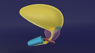
The third, most advanced, last to develop, and caudal most pair kidneys is metanephric kidney. It lies in the pelvic region and forms a mesenchymal condensation called metanephric blastema.
From the caudal end of the mesonephric duct and diverticulum arises that goes towards the metanephric blastema. As it reaches close to the blastema, in its honor, it becomes dilated and gets divided several times.
The last division forms the collecting tubule and each tubule has a small cap of metanephric blastema at its distal end. There are millions of collecting tubules, each wearing its own metanephric cap. The terminal division of the ureteric bud (the diverticulum that arose from the mesonephric duct) induces the development of metanephric blastema, while the metanephric blastema itself causes the ureteric bud to divide and differentiate. This is like a group of two best friends who motivate each other to study hard. Such relationship of inducing each other is called reciprocal induction.
Whole this structure is surrounded by a thin layer of remaining blastema that forms the capsule of this definitive kidney.
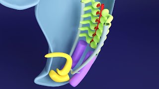
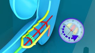
The blastemal cap forms a vesicle by process similar to that of promesonephros. i.e. mesenchyme to epithelium transition and regression of central cells to form cavity. This primitive nephron then assumes an s-shape and finally forms a full-fledged nephron. At the medial end, tuft of capillaries invaginate into it to form the bowman’s capsule. The proximal and distal part overgrows and this forms convolutions. These parts of nephron are termed as proximal and distal convoluted tubules respectively. A part of nephron beyond the distal convoluted tubule establishes connection with the collecting duct and is called connecting tubule. From Bowman’s capsule up to the connecting tubule, everything originates from the metanephric blastema while the rest of structures are derived from the ureteric bud (the diverticulum that arises from the mesonephric duct).
The diverticulum from the mesonephric duct forms the ureter. Its upper dilated part forms the basin-like pelvis of the kidney. Its first division forms the major calyx and next division forms the minor calyces.


I can't overemphasize how happy you've made me
I don't feel dumb anymore
Thank you so much. The way you demystify embryology is amazing.
after becoming dr only, i got to understand the embryology in real sense. thz alot
This is AMAZING, you are the best, please keep doing this kind of animations, it helps A LOT, love it.
Thanks a lot for your appreciation
Suggesting to all my my juniors. They are loving. It's a great help.
Thank you so much for your support
I love embryology after watching your videos❤
1:56 I think "Ureteric bud" mistakenly spelled as "Uterine bud"..
Thank u for these audio-visual 3D lectures brother... U r simply AMAZING, love u ❤️❤️
Yes you are right. It's ureteric bud. Thanks a lot for the correction and for appreciation.
Thank you for your great work and teaching!!!
you are delivering best lecturrs for medical professionals.very nice.keepit up
you are a legend
good video
Liked it. Helped very much.
Very nice presentation sir tq
Amazing 😍
Thankyou sir
its ureteric bud which develops from pronephric bud, its not uterine bud
Yes, you are right. I am sorry for the tongue slip
sooper
What is the source material or book used by you
1. Developing Human 10th edition by Keith Moore
2. Larsens Human Embryology by Gary C. Schoenwolf, Steven B. Bleyl, Philip R. Brauer, Philippa H. Francis-West
3. Langman’s Medical Embryology
4. Inderbir Singh's Human Embryology 11th Edition
5. Embryology an illustrated colour text by B S Mitchell R Sharma Robert Britton
6. Human Embryology Developmental Biology by Bruce M. Carlson 6th ed
7. Human Embryology Made Easy by Abdul Hamid Rana
8. Clinical Embryology Robert Carachi
9. Embryology by Robert E Coalson
10. Human Embryology Prenatal Development of Form and Function by W. J. Hamilton
11. Developmental biology by Michael J. F. Barresi, Scott F. Gilbert
The animations are amazing and appreciate your efforts. But the formation of blood vessels and glomerulus is bit vague and it was not described much about it. Seems all of sudden
Thank you so much for your appreciation and feedback.
You are astute in your observation that development of the blood vessels and glomerulus is quite vague. I am fully aware of this shortcoming. Unfortunately, I could not find any more details on this topic, in any of the known textbooks of Embryology. Furthermore, the research in this regard seems quite deficient. I assume that these mechanisms of embryogenesis of glomerulus have not yet been studied in adequate details. Nonetheless, I will try my best to create a separate lecture based on the journal articles that I could find on this topic.
You can always contact me for if you have relevant resources at draizaz@medicovisual.com.
Also, the development of blood vessels has been discussed in more details in this visual lecture: czcams.com/video/8wuWgDCj5u0/video.html
@@MedicoVisual Thank you for the reply. You need not to actually. But It shows how committed to your work and already you proved to be a wonderful teacher. All the best.
@@user-xk3ml3kk4z You are always welcome. Teaching and research (specifically, literature review) is my passion
7:55 🐢
Testes development proper animation plz. I am very depressed bcz am enable to understand how sex cord develop into seminiferous tubules plzzzzzz do make a video
Development of testes has been taught in a great detail in three parts visual lecture series in our "Premium 3D Urogenital Embryology MasterClass", available here: www.medicovisual.com/courses/premium-urogenital-system-3d-masterclass
Please email at draizaz@medicovisual.com or WhatsApp at +923037241030 if you need any help or have any question.
Tqqqqqqqqqqqqqqqqqqqqqq ❤❤❤❤❤
Thank you sir! Are you going to upload reproductive system embriology too?
No plans yet. It would be paid.
@@MedicoVisual okay, so you’re going to upload it on your site?
@@user-ox2gx1tr8v yes
@@MedicoVisual in the next days or we should wait a bit longer? Your courses are really helpful
@@user-ox2gx1tr8v Thank you so much for your appreciation. Urinary Bladder and Urethra embryology is recorded and will be uploaded within 2-3 days (on youtube). But genital system will take time. A few weeks maybe. I am sorry, these lectures take a long time. As I am doing everything alone.
you are a legend
Thank you so much