3D Kidney Embryology Part 1: Intermediate Mesoderm - Urogenital Ridge - Nephrogenic Cord

Vložit
- čas přidán 19. 12. 2022
- In this 3D visual lecture of Embryology, Dr. Aizaz from MedicoVisual talks about the formation of Urogenital ridge, Genital ridge and Nephrogenic cord. How the primitive kidneys like Pronephros, Mesonephros and Metanephros develop.
With the embryonic folding, along with other components of trilaminar germ layer, the intermediate mesoderm also folds. At the cranial and caudal end of the embryo, there is no intermediate mesoderm.
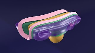
The intermediate mesoderm and the paraxial mesoderm get separated from the lateral plate mesoderm. Paraxial mesoderm goes on to form the vertebral column and contributes to the muscles and skin formation. While the intermediate mesoderm is now behind the lateral plate mesoderm. Specifically, it is behind the intraembryonic somatic mesoderm near its junction with the intraembryonic splanchnic mesoderm. This intraembryonic somatic mesoderm will form a thin epithelial membrane termed mesothelium and the epithelium is also referred to as “celomic epithelium”.
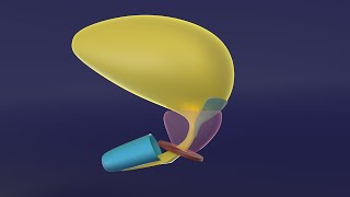
The intermediate mesoderm proliferates and forms a thick cylindrical structure called the “Nephrogenic cord” (Nephrogenic; means kidney-forming). The nephrogenic cord in its midpart is accompanied by another mesodermal mass (also derived from the intermediate mesoderm). This mesodermal mass, covered with the overlying celomic epithelium, lies slightly ventral and medial to the nephrogenic cord and is the primordium of most of the parts of gonads (testes/ovaries). Hence it is termed as genital ridge. The nephrogenic cord and ridge along with the overlying celomic epithelium form a bulge, termed as “Urogenital ridge”.
In humans, three pairs of kidneys develop in a craniocaudal fashion with increasingly advanced design.
The first pair develops in the cervical region, called Pronephros, and is the most primitive type of kidney.
Pronephros is basically a mesenchymal tissue. Mesenchyme consists of irregularly shaped cells interspersed between a ground substance. In other words, mesenchyme is the embryonic connective tissue.
The mesenchymal cells in pronephros become arranged in pair of blobs or segments called nephro-tome (NOT NephroStome) - as in sclerTome or DermoTome. Cells in the center/core of each blob die or undergo atrophy. While the cell at the periphery acquires a particular shape and establishes cell-to-cell contact. In other words these cells undergo mesenchymal-to-epithelium transition. So, this primitive pronephric nephron consists of epithelial cells lined cavity.
It then assumes a specific shape and forms an s-shaped pronephric tubule. At the anteromedial end of this tubule, a tuft a capillaries from a branch aorta gets pushed into it and forms the renal corpuscle. However, in humans pronephros are non-functional and such glomeruli do not form in human fetuses. The lateral end of the tubule joins with lateral end of the neighboring tubule immediately caudal to it. A series of such connection forms a long duct craniocaudally called pronephric (collecting) duct.
From the last pronephric tubule, the duct grows caudally downwards and along the medial side Intermediate mesoderm and even grows beyond the caudal end of this mesoderm to finally meet with the cloaca.
The pronephric kidney is the most primitive and basic type of kidney. In small fishes, it is functional and definitive kidney. Their bodies aren’t too complex and they live in water. It is very easy to excrete the limited variety of waste products formed in body of such fishes with simple type of kidney. However, humans are much more complex. They ingest and create several different type of toxic substances and have large bodies. Such simple kidneys are simply not enough for humans. So more complex kidneys are needed. The pronephric kidneys in human are non-functional and regress shortly. As the caudal pronephric tubules form, the cranial tubules are gradually lost, along with their adjacent portion of their duct.
As the cranial portions are lost the caudal most portions of pronephros induce the formation of next more advanced pair of kidneys called mesonephros. Mesonephric tubules form in a similar fashion as pronephric tubules. The tubules create urine and collecting duct is involved in transport of urine to urinary bladder and also involved in pulling water thus further concentrating the urine even. So for this primitive function there is no need to re-invent the wheel. Rather than forming its own collecting duct, it uses the distal (and remaining) part of the pronephric (collecting) duct. After mesonephric kidneys assert their ownership over the pronephric duct, now it renamed as mesonephric duct, also called Wolffian duct on the name of scientist who first described it.
Even the mesonephric kidney is not enough to cater the needs of an organism as complex as human. So most of the tubules of mesonephros regress along with their adjacent collecting duct. However, in some smaller animals, mesonephric kidneys are the definitive kidney.


The worth of watching a 30 minute lecture to actually understand embryology is such a bliss
Thank you. Unlike my professors, you don't go around showing off your prizes and stuff you've accomplished I don't care a damn about. You actually TEACH EMBRYOLOGY!
The way you explain embryology is amazing!!! These are the best videos to make your embryology concepts crystal clear !!!
After much effort, I have found this video...
But really, I have understood everything now that I didn't understand from other videos...
Thank you very much for this kind of animation with much explanation
Please upload remaining part also.
Really helpful 👍👍👍
i've been struggling with the concepts so much and i finally found the answers, thank you so much!
First time in my life embryology seems interesting and all credit goes to ur video ❤❤❤
thx allllllooooot you are amazing 🎉🎉🎉🎉🎉
Thank you sir!!
Your work means a lot and helps clear our understanding
These videos are so helpful, I love them
Amazing❤❤❤❤❤❤❤❤
One of the best explanation 👌
WONDERFUL, THANKS A LOT SIR
thank u SOSOSOS much wow
Excellent, Sir...... ❤
Wow.. Great effort
thank you sir very much!!!! for me you are a blessing of allah almighty...may allah bless you more than you think...❤
Thank you so much
❤️❤️❤️
Sir u are one of the best.
The best 🎉
LOVE YOUUUUUU!!!!!!
Thank you so much ❤
Fantastic
Wonderfull
Amazing
Gooooood job buddy
Thank you sir 👏👏👏👏 l appreciate the effort
Wish we had this during mbbs
sooper sir
BEST ❤
You are god sent 😍
you save my life
zabardast
Great vid !
Thanks alot
+Do you design these 3D models by yourself?
Always welcome.
Most of the 3D models used in my visual lectures are created by me in Blender 3D software. However, some were bought from websites like turbosquid, sketchfab, and free3d.
Lengthy but worth watching
Good things take time 😉
You are amazing Doctor and excellent tutor sir
Can you please make videos in genital system development please 😢❤?
Hi.
Thank you so much for your appreciation.
Genital System Embryology is already available for a small fee at www.medicovisual.com/courses/premium-urogenital-system-3d-masterclass
Do not hesitate to contact me if you have any questions or concerns.
WhatsApp: +923037241030
Email: draizaz@medicovisual.com
What program or app did the Dr. used to show us the embryo?
Blender 3D (www.blender.org)
hi, i wonder if you can tell me what platform or programme do you use for the resources, i need to study a lot for embriology
Hi. I am sorry for the late response. I have been busy lately.
I start with a basic medical embryology book. Typically BRS, if I already have some understanding of the topic, or Langman if the topic is new for me.
I then study it from multiple books of Embryology. Here are my top-picks:
1. Developing Human 10th edition by Keith Moore
2. Larsens Human Embryology by Gary C. Schoenwolf, Steven B. Bleyl, Philip R. Brauer, Philippa H. Francis-West
3. Langman’s Medical Embryology
4. Inderbir Singh's Human Embryology 11th Edition
5. Embryology an illustrated colour text by B S Mitchell R Sharma Robert Britton
6. Human Embryology Developmental Biology by Bruce M. Carlson 6th ed
7. Human Embryology Made Easy by Abdul Hamid Rana
8. Clinical Embryology Robert Carachi
9. Embryology by Robert E Coalson
10. Human Embryology Prenatal Development of Form and Function by W. J. Hamilton
11. Developmental biology by Michael J. F. Barresi, Scott F. Gilbert
For most of the topics, just knowing Embryology is not enough. To understand clearly, I also go through relevant topic from textbooks of Anatomy, Histology and Physiology.
Regarding the animations, I mainly use "Blender" it is free and open-source. For 2D, PowerPoint is enough.
Hai sir . Iam not a medico but I like to know embryology. I appreciate ur work.
Thank you for your kind words! I'm glad to hear that you are interested in embryology. Even if you are not a medico, you can still learn a lot from these visual lectures. Feel free to ask any question that you may have. 😊
Thank you for the amazing work, you definitely have to power of being crystal clear and simple!
Sorry to bother you, but I was wondering where do the gonads come from. I understand that female + male ducts originate respectively from paramesonephric and mesospheric duct, but as far as I understood from the Langman textbook the gonads are just formed medially (even medial to the mesonephric kidney), so that it seems that they should originate from the genital cord you mention in the beginning of the video. But the genital cord is not mentioned in the Langman.
Thank you so much for your appreciation.
According to several authors (I am sorry I don't remember the exact reference but one of the book that I can remeber is "Netter's Atlas of Human Embryology"), nephrogenic cord is the mesenchyme that underlies the bulge into posterior abdominal wall. This bulge is called Nephrogenic "ridge". In other words, celomic epithelium + underlying mesenchyme = ridge.
Now in literature, you will almost always find, Nephrogenic cord and Genital ridge (and not the Nephrogenic ridge and Genital cord). This is because gonads are formed by both, the mesenchyme and the celomic epithelium while kidneys are only formed by underlying mesenchyme (cord part only).
This is not a proper theory or a rule, its my observation after reading this topic from 20+ textbooks and some journal articles. Some book still arbitrarily use these terms interchangeably. I really wish there should be a central body of anatomists/embryologists to properly define and enforce a standardized usage of terms. (Please let me know, if there is already)
Where does the genital ridge come from?
Most authors agree that genital ridge develops as result of proliferation of mesenchyme of intermediate mesoderm and to some extent the overlying epithelium. In other words, the intermediate mesoderm proliferates to form two types of buldges, the nephrogenic part and gonadal part. In essence, both have similar origin.
I hope I was able to clear your doubts.
Also please note that as I mentioned in my previous comments, these terms are not standardized, unfortunately. So some textbooks like Keith More's Developing Human even use the term genital cords to refer to the primary sex cords. Things become really confusing this way.
@@MedicoVisual It was very helpful, thank you!
What the app you using for this?
Please answer i need the app
It's a software called Blender
I guess everyone who studies embryology needs your premium subscription. Do you put videos that you will put later in the future, or is it necessary for us to buy the premium masterclass course to get the full content?
Thank you for reaching out with your question! 🌟
At MedicoVisual, we offer a mix of free and premium content.
While most of our system-based Embryology videos are part of the paid subscription, we also have free offerings, such as our Ear Embryology course, available on CZcams and our website for everyone. Furthermore, some videos from our paid courses are also available for free on CZcams (like this one).
Our premium subscription includes additional resources like MCQ-based quizzes for comprehensive practice.
If you're interested in specific systems, you can purchase them individually, like CVS Embryology or GIT Embryology. We also provide a bundled option with all courses at a discounted price.
We're committed to making our content accessible, so if the cost is a concern, please feel free to contact me at draizaz@medicovisual.com or via WhatsApp at +923037241030 for a personalized discount.
We believe in supporting all students on their medical journey! 😊📚
@@MedicoVisual that is a very detailed and useful answer, thank you
@@beinghimself Thank you so much.
My students are always my top-priority 😊
From where coelomic epithelium is derived??
Lateral plate or intermediate mesoderm??
Thanku sir❤
Lateral Plate Mesoderm; specifically Somatopleuric Part of Lateral Plate Mesoderm.
❤️❤️❤️❤️❤️
Kindly upload the urogenital system embryology
Urogenital System Embryology is already available for a small fee at www.medicovisual.com/courses/premium-urogenital-system-3d-masterclass
Do not hesitate to contact me if you have any questions or concerns.
WhatsApp: +923037241030
Email: draizaz@medicovisual.com
❤❤❤🎉🎉🎉😊😊😊
Woowww waiting for this😭😭😭😭😭😭 thank you sirr this will help me a lott
Always welcome. Thank you so much for your support.
Please , give us the pdf if you have 🎉 .
I am sorry. PDF Notes are not available as of now. It is a priority though.
@@MedicoVisual or even illustrations ?
@@dr.ahmedal-yousif7398 Illustrations will be included in the PDF notes
@@MedicoVisual I will stay waiting you 😍😍 .
Thanks I appreciate what u offer... But u gonna on and on repeating without clear points... Leaving us in a circle without jumping on next one... Anyway thanks
I am sorry that you do not like my style of presentation. Please watch all parts in series. That might make more sense and help you better understand. I truly appreciate your constructive criticism. Looking forward to hear more from you. Criticism helps a lot in improving my lectures.
Also, can you please share some insights on how can I improve it?
This is so true
@@MedicoVisualyou don't have to overemphasize on things you've said before..if we didn't get it the first time we could go over the video again
The repetition is weakening but again thank you so much❤
Hello @@chibuzornoble6442
Thank you for your honest feedback. I appreciate you pointing out that repetition can be overdone. I aim to cater to students at different levels, but I understand the need to strike a better balance. I'll keep your suggestion in mind for future videos to ensure they're concise and beneficial for all viewers.
Thanks again for your honest feedback and for helping me improve. Your input is invaluable!
Best regards,
Dr. Aizaz from MedicoVisual