Photoacoustic tomography: ultrasonically breaking through the optical diffusion limit

Vložit
- čas přidán 6. 08. 2024
- Lihong Wang's Hot Topics Presentation from SPIE Photonics Europe.
spie.org/photonicseurope -
Photoacoustic tomography: ultrasonically breaking through the optical diffusion limit
Lihong V. Wang, Optical Imaging Lab., Washington Univ., United States
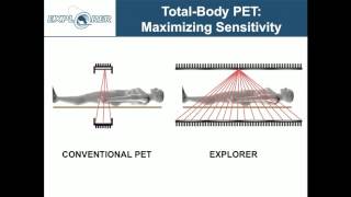
Photoacoustic tomography (PAT), combining optical and ultrasonic waves via the photoacoustic effect, provides in vivo multiscale non-ionizing functional and molecular imaging. Light offers rich tissue contrast but does not penetrate biological tissue in straight paths as x-rays do. Consequently, high-resolution pure optical imaging (e.g., confocal microscopy, two-photon microscopy, and optical coherence tomography) is limited to depths within the optical diffusion limit (~1 mm in the skin). Ultrasonic imaging, on the contrary, provides good image resolution but suffers from poor contrast in early-stage tumors as well as strong speckle artifacts. In PAT, pulsed laser light penetrates the tissue and generates a small but rapid temperature rise, which induces emission of ultrasonic waves due to thermoelastic expansion. The ultrasonic waves, ~1000 times less scattering than optical waves in tissue, are then detected to form high-resolution images at depths up to 7 cm, breaking through the optical diffusion limit. Further depths can be reached by using microwaves or RF waves as the excitation source. PAT, embodied in the forms of scanning photoacoustic microscopy or photoacoustic computed tomography, is the only modality capable of imaging across the length scales of organelles, cells, tissues, and organs with consistent contrast. Such a technology has the potential to enable multiscale systems biology and accelerate translation from microscopic laboratory discoveries to macroscopic clinical practice. PAT may also hold the key to the earliest detection of cancer by in vivo label-free quantification of hypermetabolism, the quintessential hallmark of cancer. The technology is commercialized by several companies.
Biography: Lihong Wang earned his Ph.D. degree at Rice University, Houston, Texas under the tutelage of Robert Curl, Richard Smalley, and Frank Tittel. He currently holds the Gene K. Beare Distinguished Professorship of Biomedical Engineering at Washington University in St. Louis. His book entitled "Biomedical Optics: Principles and Imaging," one of the first textbooks in the field, won the 2010 Joseph W. Goodman Book Writing Award. He also coauthored a book on polarization and edited the first book on photoacoustic tomography. Professor Wang has published over 280 peer-reviewed journal articles with an h-index of 57 and delivered more than 310 keynote, plenary, or invited talks. His laboratory invented or discovered functional photoacoustic tomography, 3D photoacoustic microscopy (PAM), the photoacoustic Doppler effect, photoacoustic reporter gene imaging, focused scanning microwave-induced thermoacoustic tomography, the universal photoacoustic or thermoacoustic reconstruction algorithm, frequency-swept ultrasound-modulated optical tomography, time-reversed ultrasonically encoded (TRUE) optical focusing, sonoluminescence tomography, Mueller-matrix optical coherence tomography, optical coherence computed tomography, and oblique-incidence reflectometry. In particular, PAM broke through the long-standing diffusion limit to the penetration of conventional optical microscopy and reached super-depths for noninvasive biochemical, functional, and molecular imaging in living tissue at high resolution. His Monte Carlo model of photon transport in scattering media is used worldwide. He has received 31 research grants as the principal investigator with a cumulative budget of over $34M. Professor Wang is a Fellow of the AIMBE (American Institute for Medical and Biological Engineering), OSA (Optical Society of America), IEEE (Institute of Electrical and Electronics Engineers), and SPIE (Society of Photo-Optical Instrumentation Engineers). He is the Editor-in-Chief of the Journal of Biomedical Optics. He chairs the annual conference on Photons plus Ultrasound, and chaired the 2010 Gordon Conference on Lasers in Medicine and Biology and the 2010 OSA Topical Meeting on Biomedical Optics. He is a chartered member on an NIH Study Section. Wang serves as the founding chairs of the scientific advisory boards for two companies commercializing his inventions. He received NIH's FIRST and NSF's CAREER awards. He was awarded OSA's C.E.K. Mees Medal and IEEE's Technical Achievement Award for "seminal contributions to photoacoustic tomography and Monte Carlo modeling of photon transport in biological tissues and for leadership in the international biophotonics community". - Věda a technologie


Thank you Prof. Wang, that was a video, that persuaded me to start with optoacoustic imaging, and that was about 8 years ago!
I am high school student form Turkey and I am preparing a research paper on this topic, mostly on photoacoustic effect and phostoacousic imaging. This presentation was incredibely beneficial for me. Hoping to see photoacoustics more on future.
This is absolutely unbelievable. I had no idea this imaging modality even existed. incredible work.
Thank you for posting these. It is cutting edge work, amazing we can view for free here.
I participated in Photoacoustic research at the University of Massachusetts in Boston during my undergraduate years. I will be giving a presentation on photoacoustics in a few days, and this seminar has been very helpful.
There are many great applications for photoacoustics, and I am excited to see what the next twenty years will bring. Thanks!
Can you share the presentation (with animations) on photoaccoustic?
Thank you so much for uploading. This is my interested tech.
Indeed it is, the field is growing quickly. The conference at our Photonics West Symposium has grown exponentially each year since its introduction. Keep an eye out, we will be publishing an interview with Lihong Wang in the next week or two.
Where can i get ANSI standard for academic purpose, Thanks in advance.
Very very great talk!
41:33 Regarding scanning through thick bone. If you'll forgive the simplification, PAT seems like a type of camera which can effectively 3D scan the shape of it's own lens, but in this case the lens being flesh and bone. So can't you use the accumilated scan data from shallower layers as a waveguide/mask to virtually deblur deeper layers?
i imagine the scanning process being a raster which gradually goes deeper and deeper, layer by layer. The data from each deeper layer goes through a sort of lens-correction filter, effectively put through a software "virtual lens" made up of the accumilated data from the previous layers.
A bone may blur your image, but you have the capability to scan the exact structure of the bone, thus you have knowledge of the exact nature of the blurring.
Very interesting. I like how the presenter uses "we used math" in the same way one says "we used magic"
Can I get the data sets for the PAT
This looks highly interesting. Unfortunately, something has happened to the sound, making it barely audible and filled with odd clicks.
This is odd. I can hear any other YT video just fine, but this one is so faint that I can barely make out the words, and word endings sometimes have a weird squelch. I went to your website, and the audio is slightly louder, but I still have to have my speakers turned up all the way to hear what is said.
I just listened to the presentation and did not have the same problems. please try again, also the video is posted on our website - spie (dot) org/x86922.xml
I just swapped out my new speakers for my old, defective speakers. As I suspected, the volume plays just fine on my old, defective speakers.
I suspect the problem has to do with the channels used in the sound system. Something isn't coming through the difference in design of my new speakers.