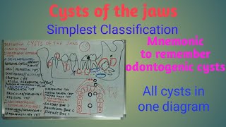
Doctor I have aquestion how can we differentiate between periapical lesion and cyst I mean when see it radiographically whet should first come to our mind because some times there is large periapical lesion
Dear Dr. Alshiky, Thank you for the question. I am assuming you are asking how to differentiate between a periapical cyst and periapical abscess/granuloma on a radiograph. When small, it is difficult to differentiate. If the lesion is about a centimeter or larger, look for the border of the lesion. If you see a corticated margin (thin white line), this is more likely to be a cyst. The margin of the abscess is usually is irregular, and surrounded by areas of sclerosis. The sclerotic bone does not have a defined margin. You will have difficulty identifying the limits of sclerosis. So, if you’re struggling to identify the extent of sclerosis, most likely it is an abscess or an infected cyst. What I have described is more of an academic debate. For patient care, it does not matter much whether the lesion is an abscess or a granuloma or a cyst. This tooth needs to be treated, either endodontically or surgically. Since the treatment is the same, I would prefer to call a smaller lesion as apical periodontitis and recommend the same treatment. In the periapical area, sometimes odontogenic keratocyst may also happen. Make sure that you can differentiate between these two lesions. If the tooth is vital and there is a periapical lesion, this may be odontogenic keratocyst. Typically, a keratocyst is tunnel-shaped while a radicular cyst is circular/oval. Earlier I had posted a video (czcams.com/video/xkdpodba1JI/video.html) of a keratocyst that almost looked like a radicular cyst. That video might help you with your question. I hope this explains.


One of the best videos ever made for dental students 😊
I’m your Number one fan Dr. Ahmad, keep on rockin in the free world!
Hey, Tevin! You are so kind, as usual!
You are God's gift to every dental professional. Thank you so much.
Wow! Thanks!
Your videos are beyond praise , too good
Cannot justify with words .
They are brief to the point yet detailed but not too much detailed, just right!
Quality of this channel is something to behold
Dr. Oflaz, you are so kind!
All videos channel is watched
Saturday 23/1/2020🖤🖤🖤
I’m from iraq 🇮🇶 specifically kufa city
Big thanks 🙏 ♥️♥️
Dr. Adil, So glad that you took the time to watch all the videos. Hope these were helpful.
Preparing for Oral Surgery boards. I've found this to be an amazing resource.
Amazing
Perfect explanation 👏🏽thx a lot
A fantastic lecture sir !!
Most welcome!
Should i go to a dentist if i suspect to have a cyst in the jaw?
Great explanation you are the best one describe oral radiology keep going🙏🏻
Great sir keep it up 👌
Dr. Najeeb Ullah, Thank you for stopping by and your comment.
I have also same problem
Cristal clear sir.... thank u
Thank you, Dr. Aggrawal!
could you help me? I have this problem and if you have time I would share the 3d image
Thanks for that! A polycystic, enlers-danlos syndrome !!! I was blessed with cysts making! I hate my body!
🍁
Doctor I have aquestion how can we differentiate between periapical lesion and cyst I mean when see it radiographically whet should first come to our mind because some times there is large periapical lesion
Dear Dr. Alshiky, Thank you for the question. I am assuming you are asking how to differentiate between a periapical cyst and periapical abscess/granuloma on a radiograph. When small, it is difficult to differentiate. If the lesion is about a centimeter or larger, look for the border of the lesion. If you see a corticated margin (thin white line), this is more likely to be a cyst. The margin of the abscess is usually is irregular, and surrounded by areas of sclerosis. The sclerotic bone does not have a defined margin. You will have difficulty identifying the limits of sclerosis. So, if you’re struggling to identify the extent of sclerosis, most likely it is an abscess or an infected cyst. What I have described is more of an academic debate. For patient care, it does not matter much whether the lesion is an abscess or a granuloma or a cyst. This tooth needs to be treated, either endodontically or surgically. Since the treatment is the same, I would prefer to call a smaller lesion as apical periodontitis and recommend the same treatment. In the periapical area, sometimes odontogenic keratocyst may also happen. Make sure that you can differentiate between these two lesions. If the tooth is vital and there is a periapical lesion, this may be odontogenic keratocyst. Typically, a keratocyst is tunnel-shaped while a radicular cyst is circular/oval. Earlier I had posted a video (czcams.com/video/xkdpodba1JI/video.html) of a keratocyst that almost looked like a radicular cyst. That video might help you with your question. I hope this explains.
I appreciated as usuall clear and pricise explantation thx
21:55 residual cyst
مافيش برامج طبية عن cyst