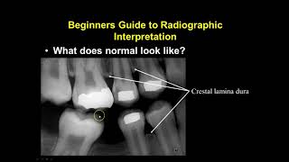
Radiographic Interpretation of Pulp and Periapical Infections। Chairside diagnosis

Vložit
- čas přidán 5. 09. 2024
- This video talks about the distinct radiographic changes in the following lesions and helpful for final year BDS exams:
1. Chronic Pulpitis
2. Acute Apical Periodontitis
3. Acute Periapical Abscess
4. Chronic Periapical Abscess
5. Chronic Apical periodontitis (Periapical Granuloma)
6. Periapical Cyst
7. Periodontal Abscess
8. Endo-Perio Lesion
9. Condensing (Sclerosing Osteitis)
10. Infected Peripical Cyst
To know more, watch the video..


A big thanks for. This mam. I was never able to understand this concept before this.. but now everything is crytal clear
Maám i watched this video two times a day,,
1st i watched ur stunning expression toward teach,,❤️
2nd time i understand purpose of video 😊
You explained each and everything so clearly and in a simple way. Thanks 👍
My college oral Medicine and Radiology staffs teaching about Peri apical cyst well defined radiolucecy surrounded by sclerotic border. Not a corticated border. Nice presentation madam.
Thank you..n U have to follow what is taught in ur college in the end..actually there is some variation in radiographic diagnosis in all the colleges..actually at BDS level, terms sclerotic and corticated are used synonymously..the difference u can see in white and pharaoh..that's what I have told in my video u need to follow what the staff teaches you in the end..
Thank you so much ma'am.👍👍
Hello Shilpi mam..Your video is awesome ..❤loved the way you cleared each and every term😘
So helpful.. thank you so much ♥️
Very helpful video. Thank u for sharing
Awesome Video mam thank u so much really needed this as a reference for my OMDR posting.
Thank you so much..
Thanks a lot Maam ....you have solved my problem......hope to see more videos based on odontogenic cyst and tumor
Very knowledgeable video thanks mam for this information
Thank you so much..😊
Dr Shilpi, you are beautiful!
Please smile in the future photos 😁
It was a great video ma'am.
Please tell how to determine size of a lesion approximately with the help of a radiograph
This video is so helpful.
In acute apical periodontitis there is no break in continuity of lamina dura...only pdl space gets widened...if break in continuity of lamina Dura also present then it's suggest the presence of periapical abscess
Thank you for the video , please do video on cicatrization of periapical lesion, bone, dentin and ALD ..
Nicely explained madam
Great 👍
Nicely explained 👌
thank you very much!!
Superb Class Madam.... 👍🏼👍🏼💓
Thank you..
Thank you 👏🏻👏🏻👏🏻
thank you for the video Dr
Love the way u teach
Thank you so much..
Excellent informative video. From 🇵🇰
Thank you so much..❤️
@@fortheloveofteaching-drshi5011 keep shining.
I like the way you teach😊😍
Thank you so much..
Very helpful 👏
You need digital board
Btw your explanation excellent ✌️🎉
Joss
Very nice Dr
Thank you so much doctor..😊
What is the diagnosis when laterally to an upper lateral incisor with post core and crown there is huge radulucency ?
Love yr teaching 🥰❤️
Thank you so much..
Very nicely explained 👌
Thank you sir..
Explain thanks ma'am
Maam, if there is only loss of lamina dura then what is the diagnosis.
❤❤❤
Thankyou!! Very helpful☺
Thank you so much..
Thank u informative
Thank you so much..
Great 👏🏼👏🏼
Thank you..😊🙏
Happy Teachers Dayy✨️✨️❤️❤️
Thank you..
How sclerotic border differs from corticoid border
What is the difference between periodontal abscess and periapical abscess?
Thanks you keep up 🙂🙂
Thank you so much..
Hi doc, that's a nice presentation, however the classification that u have mentioned here does not follow the AAE guidelines for clinical classification of endodontic diseases.
Thank you..I haven't classified anything in the video I guess..anywaz can u please share the same with me? it would be ready helpful..
Thank you ! could you make video on types of osteomyelitis and it's radio features?
Yes..for sure..
I love you your teching
U must discuss the management too
Oh..k..sir..👍
Thank you madam
Thank you so much..😊
Ma’am didn’t get the basic difference in the diagnosis of Acute peri apical abscess and acute apical periodontitis. Found both of em sorta similar
Yes..radiographically they are similar only..as the bone loss is not significant in acute abscess to see changes in the radiograph..to agar apko radiographically widening of PDL space with ou without loss of lamina dura dikhta h..to ap AAP radiograph diagnosis denge..final diagnosis ap clinically relate karke denge..but jab abscess me kisi chronic lesion me acute infection ho jata h..tab apko well defined lesion me kahin kahin borders p fuzziness dikhne lagegi..
But apke college me apko Jo bhi bataya jata h vo follow karna h...kunki har college me thode thode changes hote hain..agar ap concept samjhenge to apko bohot easily samajh ayega Jo bhi vo kehna chahte hain..apko koi doubt h to puch sakte hain..
@@fortheloveofteaching-drshi5011 Thank you ma'am , I'm much more comfortable with it now compared to what it was before! ❤️🙏
mam please meri help karie mere pore mouth me pata nahi kya ho gaya hai white white si line hai or hot suje hue hai mujhe aapko picture bhejni hai plese mam madad karie
Hello mam perio-endo lesion, endo- perio lesion both are same right??...
Yes it is..but the terms are used differently sometimes..perio endo means primarily a perio lesion leading to secondary endodontic involvement...and endo perio means primarily and endo lesions leading to secondary periodontal involvement..
👍
Hindi me smjh me aata h madam
❤u👍
Thank you mam 🙂
😢
:)