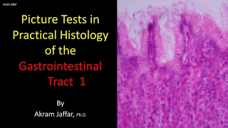
Picture tests in histology of the gastrointestinal system 5

Vložit
- čas přidán 23. 02. 2017
- After completion of this video you will be able to:
Differentiate between: contents of a portal tract, appendix and remaining large intestine, small and large intestine
Describe: Paneth cell: location and function, blood flow in the liver, function of colonic mucosa, location and function of submucosal plexus.
Identify: enterocyte, goblet cell, villi, crypts, circular folds (plicae circulares), Paneth cell, Brunner’s glands, lymphoid follicles, portal tract, centrilobular venule, hepatocyte, hepatic sinusoid, hepatic cord.
Presented and edited by Dr. Akram Jaffar, Ph.D.
This video and its channel are supported by "Human Anatomy Education" Page on Facebook / anatomyeducation
Subscribe to the channel to receive updates.
Feedback is highly appreciated from channel viewers
Some images, with gratitude, were cited in:
www.researchgate.net/publicat...
faculty.cord.edu/todt/336/lab/...
www.histologyguide.org/index.html
www.siumed.edu/~dking2/erg/liv...


but there says duodenum....
??