Dural Folds | Falx cerebri | Tentorium cerebelli | Falx cerebelli | Diaphragma sella | Attachments |

Vložit
- čas přidán 5. 05. 2020
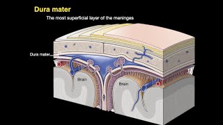
- The dura mater is the outermost, thickest and toughest membrane covering the brain
There are two layers of dura:
a. An outer or endosteal layer which serves as an internal periosteum or endosteum or endocranium for the skull bones.
b. An inner or meningeal layer which surrounds the brain. The meningeal layer is continuous with the spinal dura mater.
The two layers are fused to each other at all places, except where the cranial venous sinuses are enclosed between them.
Endosteal Layer or Endocranium:
1. The endocranium is continuous:
a. With the periosteum lining the outside of the skull or pericranium through the sutures and foramina.
b. With the periosteal lining of the orbit through the superior orbital fissure.
2. It provides sheaths for the cranial nerves, the sheaths fuse with the epineurium outside the skull. Over the optic nerve, the dura forms a sheath which becomes continuous with the sclera.
3. Its outer surface is adherent to the inner surface of the cranial bones by a number of fine fibrous and vascular processes.
The adhesion is most marked at the sutures, on the base of the skull and around the foramen magnum.
Meningeal Layer
At places, the meningeal layer of dura mater is folded on itself to form partitions which divide the cranial cavity into compartments which lodge different parts of the brain.
The folds are:
1. Falx cerebri,
2. Tentorium cerebelli,
3. Falx cerebelli,
4. Diphragma sellae.
Falx cerebri
The falx cerebri is a large sickle-shaped fold of dura mater occupying the median longitudinal fissure between the two cerebral hemispheres.
It has two ends:
1. The anterior end is narrow, and is attached to the crista galli.
2. The posterior end is broad, and is attached along the median plane to the upper surface of the tentorium cerebelli.
The falx cerebri has two margins:
1. The upper margin is convex and is attached to the lips of the sagittal sulcus.
2. The lower margin is concave and free.
The falx cerebri has right and left surfaces each of which is related to the medial surface of the corresponding cerebral hemisphere.
Three important venous sinuses are present
in relation to this fold.
1. The superior sagittal sinus lies along the upper margin;
2. The inferior sagittal sinus along the lower margin
3. The straight sinus along the line of attachment of the falx to the tentorium cerebelli
Tentorium cerebelli
• The tentorium cerebelli is a tent-shaped fold of dura mater, forming the roof of the posterior cranial fossa.
• It separates the cerebellum from the occipital lobes of the cerebrum,
• broadly divides the cranial cavity into supratentorial and
infratentorial compartments.
• The infratentorial compartment, in other words, is the posterior cranial fossa containing the hindbrain and the lower part of the midbrain.
• The tentorium cerebelli has
• a free margin and
• an attached margin
• The anterior free margin is U-shaped and free.
• The ends of the 'U' are attached anteriorly to the anterior clinoid
processes.
• This margin bounds the tentorial notch which is occupied by the midbrain and the anterior part of the superior vermis.
• The outer or attached margin is convex.
• Posterolaterally, it is attached to the lips of the transverse sulci
on the occipital bone, and on the posteroinferior angle of the parietal bone.
• Anterolaterally, it is attached to the superior border of the
petrous temporal bone and to the posterior clinoid processes.
• Along the attached margin, there are the transverse and superior
petrosal venous sinuses.
The trigeminal or Meckel's cave is a recess of dura mater present in relation to the attached margin of the tentorium.
It is formed by evagination of the inferior layer of the tentorium over the trigeminal impression on the petrous temporal bone.
It contains the trigeminal ganglion.
The free and attached margins of the tentorium cerebelli cross each other near the apex of the petrous temporal bone. Anterior to the point of crossing, there is a triangular area which forms the posterior part of the roof of the cavernous sinus, and is pierced by the third and fourth cranial nerves. The tentorium cerebelli has two surfaces. The superior surface is convex and slopes to either side from the median plane.
The falx cerebri is attached to this surface, in the midline; the straight sinus lies along the line of this attachment. The superior surface is related to the occipital lobes of the cerebrum.
The inferior surface is concave and fits the convex superior surface of the cerebellum. The falx cerebelli is attached to its posterior part
Falx cerebelli
The falx cerebelli is a small sickle-shaped fold of dura mater projecting forwards into the posterior cerebellar notch. The base of the sickle is attached to the posterior part of the inferior surface of the tentorium cerebelli in the median plane.
Follow me in blogspot - human-anatomylessons.blogspot...


Mam u r genius 💕, u may go higher.. Plzz upload all NEUROANOTOMy... So that students prefer to here ur channel becoz Dr najeeb has not explained it fully anatomically.. Plzz notice this nd see in the matter 💕💕
Sure 😊
@@HumanAnatomyLessons 💕😍😍😘👍
You just made my studying much easier, I’m so happy I could cry tears of joy right now.
Thank you sooo much, best of wishes to you and to all people reaching for free on CZcams like you
Thank you 😊
Thank you for this. My textbook only showed a midsagittal view and a posterior view of the cranium as examples and I had no idea how it all looked or connected.
Thank U
Thank you so much, it is great to memorise the relations of dural folds ✌🏼
You are welcome!
Ungaloda videola na patha first video .....enoda college hostela ...ipa varaikum unga videoslathan anatomya padichtu varen really thank u so much mam...
So happy to hear. Thank you for supporting 😊
Best video I've ever seen... nothing can be better than this...salute ma'am
Thank you 😊
Best video for understand Dural folds in very short time.. 🤩
Keep it mam🙏🙏👍
Thanks a lot 😊
I was reading it from 2 days n unable to understand! Now I got it 😌 Thank You So Much 💗
You're welcome 😊
It's very very helpful for me to see the complete view of folds.. And your explanation along with this video is just awesome combination 👍👌👌
Great effort ma'am 🙏
Thank you 😊
fantastic mind blowing
Thank you so much 😀
excellent video,we need more videos like this
Thank you, I will
The animation is remarkably easy to understand wow🔥
Thank you 😊
after watching this i can freely miss my demo class❤❤❤
This is just a media to help u what u have seen in demo
Pls don't miss any demo
Superb 😘😘
Thanks for liking
Mam thankyou so much for your hard work !
You help us study so much
May God bless you.
Thank you 😊
Best anatomy channel on CZcams for undergraduates and postgraduates 👍👍👏
Thank you 😊
Thankyou ma'am ! That was very helpful lesson for me. it is very difficult to learn anatomy only by read books because It is important to have a 3D imagination in mind to remember the right topic forever.
It's my pleasure
Thank you so much, this was a great, informative and loaded video. Please much much much love
You are so welcome!
Thank you it was really helpful
Was facing difficulty in understanding without 3d but you made it easy ❤❤❤
Most welcome
Keep watching 😊
Just wonderful😍very well explained....Thank u mam...U made learning the topic so easy....Plz go ahead mam and make more videos like this.....Thank u soo much mam...🙏
Thank you, I will
Thank you so much... You just made this easy for me.
You are so welcome!
Wow Just Awesome 🔥
Thank you so much
loved it. thank you
You are so welcome!
Wow 😲 , Just great 👍
Thank you 😊
Really helpful, thank you very much!
Most welcome
Thank you so much, ma'am!!!
Most welcome
Thank you this really cleared my doubts!please make more videos on head and neck😊
Thank you, I will
Well explained ! 🙏🙏✌🏼✌🏼✌🏼
Glad you liked it
Amazing
Thank you
Best explanation 👏👏👏
Thank you 😊
Crystal clear concept💜
Thank you
The view is amazing and easy to understand mam❤️.
Thank you 😊
Very nice presentation mam
Thanks 🙏 for great content
Thank you 😊
Thank You So Much Mam for making this easy for us
Most welcome 😊
Thanks for the clear explanation🌌
Most welcome
Thank u so much for vedio 🥰🥰👍🏼👍🏼
My pleasure 😊
Excellent ❤
Thanks 😊
finally understood arrangement of tentorium cerebelli👍🏻
Great 👍🏻
Great explanation mam.. thanks a lot
Most welcome 😊
It is very well explained. Thankyou maam
Most welcome 😊
i can't express my love for your efforts!!!...... literally you're a life saver!....God bless u
You are most welcome
Thank you so much
You're most welcome
SKULL 💀 FALX
A very nice presentation
Thanks a lot
Awesome
Thank you 😊
Best way to explain👍🏻
Thanks a lot 😊
Great explanation 🤞
Thanks 🙂
Thanks for the class
Most welcome
Sooper
Thank you 😊
Good explanation
Thank you 😊
Well explained and wonderful animation 💯
Thank you so much 😀
Nice video can u make more videos like this in head & neck part
Sure 😊
Brilliant. I am OLD and am preparing for a PG exam. It's NOT easy to study after 2+ decades have passed since I studied these basics last time and back then...I just memorized them and didn't understand much. I consulted a few sites and a few literatures to study the tentorial structures, but I just couldn't, but your lecture with video animation made it very clear.
Thank you very much for your time and effort.
Thank you 😊
Really amazing mam...thank you!!!!!!!
Most welcome 😊
thank you
Most welcome 😊
Thank u so much mam... It's an amazing video.... Keep it up👏👏
Most welcome 😊
Very nice one madam
Thanks a lot
Falx cerebri 6:17
Tentorium cerebelli 8:48
thanks alot
Most welcome 🙏
awesome lecture 👍👍👍
Thank you 😊
been tryin to create a mental picture of these fold for 3 days. Now can say that i made it
Happy to hear 👏👏👏
Well explained 👍👍
Thank you 🙂
keep uploaading more videos of 2 year
Thank you so much. It became so easier with the animation. Dhanywad.🙏
Most welcome 🤗
Thank you so much ma'am 💕💕
Most welcome
Thankyou.
Most welcome 😊
Too good 👍
Thanks a lot 😊
Dear Madam speaker your presentation was wonderful. Thank you
Thank you 😊
Thankyou soo much maam🙏🙏🙏
Most welcome
Madam superb explanation mam.
Thanks a lot
Thank you thank youu soooo much mam ♥️
Most welcome 😊
Very nice explanation.
Please mam do videos on upper, lower limb too please mam..
Okay sure
The fold of durameter which is ventrally related to corpus callosum is ????
corpus callosum is related to the inferior border of Falx cerebri
👍
Thank you so much mam💞
Most welcome
Thank you so much ,this is so clear and helpful
Most welcome 😊
Thank you mam
Most welcome 😊
Wonderful maam.. This was a terrible topic for me.. U made it so easy to understand❤❤
Thank you 😊
Thank u ma'am 💓
Most welcome
Thank u mam
Most welcome
thanku maam
Most welcome!
Thank you so much mam... It is so difficult to imagine all this... Thank you
Most welcome 😊
The best video on dura matter hands down
Thank you 😊
Tq very much mam...it's soooopppeeerrr
Thank you too
Thankyou so much mam
Most welcome 😊
Thankyou so much mam! :)
Most welcome 😊
Thank you ma'am
Most welcome 😊
Ma'am I hope you continue doing such videos. This was very helpful for me. Quality education for free - great service Ma'am.
Sure, will continue to do !!!
Can you please tell name of layers involved in suturing of cerebral part wounds
Can u pls specify?
do u mean intracranial surgery?
@@HumanAnatomyLessons yes please tell name of layers
Maam which app do you use for the 3danatomy ?
its 3d4 complete anatomy
Thankyou maam..
Welcome 😊
Thank you so much mam🥰🥰
Most welcome 😊
As clear as glass.
Thank U
Had to speed it up but well presented nonetheless! 👍
Thank you
Please same vedio in hindi
Hello
I need help to give file of mcq from your college please 🙏
I am from iraq
What app is this 3d illustration
3d4 complete anatomy
3d views .. 👍🏻
Thank u
whats the app name😊
It's 3d4 complete anatomy
Ma'am,
Playlist name!
Pls check the channel page, you will have playlist option
@@HumanAnatomyLessons
Yep ma'am
I found it
@@HumanAnatomyLessons
Thanks a lot for the for the work ma'am
Keep on going 💐
Most welcome
DURA MATTER