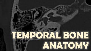
Imaging of the TMJ

Vložit
- čas přidán 22. 07. 2024
- This video describes the principles of the temporomandibular joint (TMJ) imaging, the diagnostic criteria of osteoarthritis of the TMJs, and several other disorders of the joint. This lecture uses several CBCT scans, panoramic radiographs, and MRI showing examples of TMJ disorders.


Lovely presentation!! Thanks
amazing presentation , i can't thank you enough
Dr. Elhamalawy, Thank you so much for the kind words.
Very informative discussion.
Very informative!
This is an excellent and very knowledgeable video, sir. I humbly request you to teach us space infections also. Your expertise and knowledge will help students like me.
Thank you very much for your comment. ~Mansur
Nice presentation sir
Amazing video. Thank you so much for sharing this.
A few questions please , if I may.
It's my understanding that the CBCT was taken in normal occlusion with the back teeth touching normally, without a bite block. Is that right?
Also, and forgive me if this is a stupid question, but what zoom level was used to take the CBCT?
Thank you
Thanks again doctor. I did not understand when you said one class 1 is considered class two on the table. Was that an error?
In the video you said that:
"The reconstructed panoramic radiograph shows that the scan was obtained with the teeth in occlusion."
When taking a CBCT to look at the condyles for a TMJ problem, do you think it is better to take the CBCT scan without using a bite block?
This is in order to see where the condylar head is located in relation to the glenoid fossa when the teeth are in occlusion.
What do you think?
Thank you
Thank youuuu❤
You are so kind!
Fabulous Sir. Learnt a lot
Glad to hear that!
The MRI pictures show the disc so clearly. The MRI I had doesn't show the disc so clearly. Any chance I can know what the protocol for the MRI you used there?
Thank you
very helpfull💙💙
Thank you, Dr. Ali!
❤ Hello, my friend, how are you? I have a question, please, what are the materials that X-rays cannot penetrate
Sir how to differentiate between osteoarthritis and RA based on CT of tmj
Sir plz do on sialography sir
My jaw deviate left while open and my face looks asymmetry I have prblem in speaking and laughing . Sir I have h pdf of cbct if you can catch my jaw prblem i will be very grateful to you plzz
Fix forward head posture, rounded shoulders, and anterior pelvic tilt