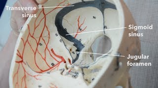
Paranasal sinuses CT imaging anatomy

Vložit
- čas přidán 7. 06. 2024
- I want to work through the anatomy of the head as seen on CT imaging sections, but there's a lot to look at. Let's start by seeing if we can recognise the frontal, sphenoid and maxillary sinuses and the ethmoidal air cells.
Scrollable computed tomography images of a normal brain:
commons.wikimedia.org/wiki/Sc...)
Paranasal air sinuses:
• Paranasal air sinuses
Music by Jahzzar
Album: HiFi City Tales
Song: Bodies
/ jahzzar - Věda a technologie


I am a radiologist, i loved this video and i think you are the best anatomy teacher of the internet.
I wish you did this same video but with a bone reconstruction CT scan. The one you are using is a soft tissue reconstruction, useful for assessing the brain in this case but the price you pay is very low quality imaging of the bone.
With bone reconstruction CT for the sinuses you can see every individual air cell of the ethmoid and how they connect with each other, every foramina, every little suture! You would love it.
Check out the radiopedia article about paranasal sinuses and look at the case examples with bone reconstruction
I wish you someday do CT / MR anatomy videos, or even a course! You would show peaople another prespective that is very useful for understanding 3D anatomy, and you be a blessing for all the radiologists.
I encourage you to try and do it, and see how radiologists all over the globe become your fans!!
Much of us would greatly benefit from the deep understanding of anatomy you provide.
Wow! So cool and I loved you explanations as to why our skull bones are the way they are. Just marvelous and super interesting!
The Bob Ross of anatomy, could listen all day 👌
Are you kidding me?! You shining the light through the bone just showed me that I'm not insane. I recently suffered a cerebrospinal fluid leak which caused intercranial hypotension and we are now in the beginning stages of trying to find the leaks location and whether it's cranial or spinal. Seeing just how sheer the facial bones are blew my mind. I'm having rhinorrhea (post nasal drip right at the back of the throat) which could also be linked. I'm just now worrying about just how delicate these bones are and how nuanced it must be to try to find leaks location/fissure with imaging. Thank you for this video, very insightful. Wish me luck trying to find my leak!
post nasal flow would indicate it is cranial, probably sphenoidal.
Contrasted CT would help
@@edwardturitwenka6115 We came to the conclusion that it's autonomic rhinorrea caused by low csf pressure from a spinal csf leak. The low pressure makes the brain sag downwards, its increased weight then puts pressure on the cranial nerves and can cause lots of strange symptoms. It is just a coincidence that both cranial and spinal csf leaks can cause a clear fluid to run from the nose. I had the fluid tested and it was negative for Beta 2 transferrin so it wasn't CSF.
Wish I had u teaching me in college years ago!!
Oh, I really like it when u do this for us , I'm from Iraq 🇮🇶, much respect for you 👏❤
Amazing !!! Thank you so much for the detailed explanation !!!!
The way u explain is awesome 😎😎😎
Love from INDIA❤️❤️❤️
Wonderful,,,, lots of respect to your teaching strategy,,, From India🙏
@samwebster You did an amazing job, breaking these down. Question that I would have loved to ask if I was sitting in your class... as far as the sphenoid and ethmoidal cavities, where would a ballon sinuplasty would be most likely to be done? Also, it would be helpful to see how they all connect to each other. Or do they?
You are my favorite … your student from Saudi Arabia 💚💚💚💚
thank you so much it's so useful and it's clearer now ! can you please talk about other anatomic structures on cerebral CT scan and also MRI ?? you made me love and understand anatomy and it helps a lot in understanding pathology and symptoms , thank you so much !!!!!!
Am a radiology resident.. u make anatomy very easy.. thanka a lot😊
Thank you for this video!!
We need more from that
Thank you ⚘⚘⚘
This was very much informative. Please do one for facial fractures too
Have you done a video on the reticular activating system? Would be great if you did! Thanks
Epic teacher! Thank you. Please also do mri axials.
I've been waiting for it soooo long 🤗
Really great video. The sinuses might be easier to see if you can get a bony sharp CT head scan.
Thank you Dr...
Thank you so much doctor
Dear doctor sam, we need more head and neck ct tutorials
I love youuuuuuuu , U SAVED MY LIFE
On its time !!! Thank you🥲🥲🥲🥲💚💚💚💚
Thank you so much
To educate people like me who have very basic human anatomy
I have question about deviated septum in the nose by birth ,
Is there any connection with
Blood pressure
I may be asking stupid question
Which laptop are you using man?
It's nice.
Quality video!!
Thank You So much !
Thank you sir.
How can I find a CSF leak in the sphenoid, ethmoid sinuses of cribiform plate?
does someone know what programme or website he is using?
Plz add subtitles to the videos and continue making videos on CT imaging
Nice 👍
Good job
But not much information Dr.)
CT is a great modality to show ostium of sinuses, osteomeatal complex and other things you can't visualisate in models
I can't breath properly,I feel a blockage behind my nose, but CT scan doesn't show anything what can that be?
pleaseee do a full head ct explaining !!! please
thanks boss
I am a medical student from iran
You are great in anatomy teaching.
What is your university name?
👏👏
it's cool, it's fun = )
It is!:)
He is making it fun for us !!
I have paranasal sinus disease. one doctor said I'm fine it's just sinuses. google search says it's cancer..
thoughts doc?
my mri side paranasal sinus disease
Warm regards from Chiajna
Hello greetings question:
1) What could be the possible diagnosis for the symptom of a foul odor emanating from one of the nasal openings "nostrils" considering the findings of a CT scan ?
2) Could the presence of mild mucosal thickening in the CT scan, potentially explain the occurrence of a bad odor originating from one of the nasal passages?
please ignore if one the questions if not apply of what you see on the CT scan result . in advance thank you
IMPRESSION: 1. Mild bilateral maxillary sinus mucosal thickening. No opacification or air-fluid level. Findings are similar to the prior CT from 11/29/2022. Signed by: mmmmmm MD 8/28/2023
Narrativa
CT FACIAL SINUSES WO CONTRAST 8/28/2023 11:13 AM AGE: 47 years GENDER: Male HISTORY: Unspecified disturbances of smell and taste COMPARISON: 11/29/2022 CT facial sinuses TECHNIQUE: Helically acquired CT of the facial bones without contrast. Axial, coronal and sagittal reformatted images were produced and interpreted. DOSAGE: Total exam DLP: 298 mGy-cm. CTDIvol (max): 24.8 mGy. Number of acquisitions at maximum CTDIvol: 1. Dose reduction was performed with automated exposure control, iterative reconstruction technique and/or adjustment of the mA and/or kV for patient size. TIMING OF STUDY WITHIN 24 HOURS OF ARRIVAL IN HOSPITAL: As outpatient FINDINGS: Metal plates and screws lining the anterior bilateral maxillary bone appears similar and uncomplicated. Old healed fracture deformities of the maxillary bone and the right and left lamina papyracea. No acute fracture identified. No findings to suggest lytic or blastic bony lesion. Soft tissue: Normal. The TMJ joints appear unremarkable. Sinuses: Minimal right frontal mucosal thickening. Mild bilateral maxillary sinus mucosal thickening. No opacification or air-fluid level. Mastoid air cells: Clear. Globes and orbits appear grossly unremarkable. Intracranial: The visualized intracranial contents are grossly unremarkable in the significant limitations of this exam not tailored for evaluation of the compartment. Dentition: No dental fracture. Cervical spine: Limited evaluation without fracture, dislocation. No significant degenerative change.
I have a sinusitis and always feel pressure from the rear of my eye because whole nasal cavity is filled with mucus😔
What's yours treatment I fell the same
12:37
Well at least we know 1 of the dislikes on this video is from Voldemort
I wish you do this with every anatomy you will save my ass I should give u half of my salary as a thank u every month since im living alone and I don’t need it other than food
You’re radiologist
Dr house
Freaking nasty.