Chest X-Ray Lung Normal Vs Abnormal Image Appearances Part 1 | TB/Pneumonia/Consolidation/Collapse

Vložit
- čas přidán 30. 06. 2024
- Chest X-Ray Lung Normal Vs Abnormal Image Appearances Part 1 | TB/Pneumonia/Consolidation/Collapse
Intro - 0:00
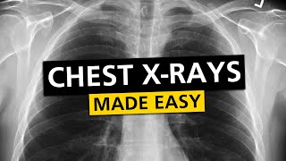
Normal Chest X-Ray - 0:07
Normal/Tuberculosis - 0:27
Miliary Tuberculosis - 2:37
Klebsiella Pneumonia - 2:52
Right Upper Lobe Consolidation - 3:47
Right Middle Lobe Consolidation - 4:16
Left Lower Lobe Consolidation - 4:36
Air Bronchogram - 4:49
Pneumonia - 5:30
Pneumocystis Pneumonia - 5:44
Pneumatocele - 6:13
Viral Pneumonia - 6:24
Right Upper Lobe Atelectasis - 6:48
Left Upper Lobe Atelectasis - 8:07
Luftsichel Sign - 8:54
Juxtaphrenic Peak Sign - 9:22
Pneumothorax - 9:36
Right Middle Lobe Atelectasis - 9:52
Left Lower Lobe Atelectasis (Retrocardiac Sail Sign) - 10:09
Double Cardiac Contour - 10:41
Flat Waist Sign - 11:03
Right Lower Lobe Atelectasis - 11:50
Superior Triangle Sign - 12:16
Consolidation/Atelectasis - 12:43


Chest X-Ray Playlist: czcams.com/play/PL4cRFWfjMmf-FXitNxxQbtOziDEL8gOON.html
Ultrasound Videos: czcams.com/play/PL4cRFWfjMmf_P02uIGRTFiYNozGKuILAX.html
Dr. Sam. I appreciate all person who use the internet to spread their knowledge. I am an old physician from Brazil. I love what internet can do as it is an important tool for a wide aspects of our society. I use it to recall and also to learn new things. Thank you very much. I really would love more if you had lateral views too. Hoping you will be able to add videos studying those images. Peace and happyness in your live and your family's.
Greetings! Thank you very much for your comment! Wishing you peace and happiness as well!
atelectasis vs consolidation of left lower lobe ; perfect mind , well discription and good job
Thank you very much for your comment! Really appreciate it!
I learned so much in your 13 mins video.
Thank you very much for watching! Glad you found it helpful!
Thank you so much!
Most Welcome!
SUPERB TEACHING
Thank you very much for watching!
Thank u
Most Welcome!
Good lecture۔Thnx for this
Most Welcome!
Thank you sir
Most Welcome!
good doc
Thanks for watching!
Doctor sir please share your impression
1. Fibrohazed densities in both upper lobes with traction of the hilum upward.
2. Diaphragm is low set and flattened with tenting of the left hemidiaphragm
3.Both costophrenic sulci are intact.
Thank you Doctor pls help me
Hello! Given these findings, a few possible diagnoses can be considered: COPD, Especially in the context of hyperinflation indicated by the low set and flattened diaphragm. Pulmonary Fibrosis or Interstitial Lung Disease, suggested by the fibrohazed densities and traction of the hilum. Post-Infectious Scarring, possibly from a previous infection like tuberculosis, particularly if the patient has a relevant history or risk factors. In some cases, chronic asthma can lead to similar findings. It's essential to correlate these findings with the patient's symptoms, history, and physical examination.
Sir mega cystitis kya hai
Report
hello doc,I just want to ask this, I need to go chest xray requirment for my nursing curriculum but I'm afraid because i have a past history of asthma and pneumonia when i was a child but its a long time ago.Will it show some problem when i get chest xray now?
Asthma typically does not leave permanent changes in the lungs that would be visible on a chest X-ray unless there has been long-term, severe disease. If you had pneumonia as a child, it is possible that there could be some scarring or changes in the lung tissue that might still be visible on an X-ray. However, this is not always the case. Past conditions, if resolved, generally do not disqualify you. Best Wishes!
Sir plz reply
No active parenchymal lesion is seen in the lung fields.
Hilar shadows are normal
CPA and apices are clear
cardiac shadows is within normal limit.
Hemidiaphragms are normal.
Bony thorax and soft tissue shadows are normal .this is
My chest xray report its normal or danger plz reply sor 0:04
Your chest X-Ray is normal
@@DrSamsImagingLibrary thank u sir