extremely well explained , very helpful , thanks a lot doctor ..... I have few questions if you allow it please : 1/- when we talk about ISNT rule in clinical exam ; do we reffer to what we see directly through a +90D lens (everything backward) or the anatomical consideration (we inverse what we see through the +90D lens) 2/- if we have an abnormality in the OCT( GCC and RNFL) with normal clinical and perimetry , treatment or not ? 2/- Could you please have the generosity to make a video explaining the interpretation of Macular OCT and fluorescein angiography. Best love and support and blessings from Algeria
As regards Q1 : yes you have to invert what you see with +90 D. Q2: OCT changes can occur in 1/3 of normal healthy persons with even small degree of myopia less than -4D. Diagnosis of glaucoma depends on +ve mutliple signs . so
for diagnosis of glaucoma you need o have more than OCT alone. The more the signs and symptoms the more one can be positive with the diagnosis Q3 : I am sorry I am not a retina doctor to cover macular OCT or fluorescein agniography in practical way. hope you will fine some other source to help with this issue.
thank you Doctor for these videos, I had read the myopia could cause the OCT for RNFL to be thinner, is it also the case for someone who's had myopia in the past but had LASIK, would it still cause the OCT RNFLY to be thinner? Thank you
@@adelabdelshafik thank you doctor, is your clinic only in egypt? so just to be clear, for myopia the possible RNFL thinning in OCT will still be the same after LASIK?
Dear Sir, can i ask, towards thr end of thr lecture, u say, upto 50%of -5DS myopes show errors of GCIPL and are fakse positive. So does is that basically mean, thst if we see a patient with apparent gcipl thinning, and fields normal and disc oct relatively normal, we should just monitor? And not treat?
The point to keep in mind is changes in OCT may occur in 1/3-1/4 of normal persons. So to diagnose glaucoma you need to have multiple different areas starting from family history, suggestive symptoms, abnormal angle configuration, suspicious disc, suspicious field changes, lower systemic BV, high myopia...... etc the more the abnormal factors the more the diagnosis is correct. so as you said, if not sure just keep pt under observation.


I'm an optometry student from Melbourne, I find your video extremely helpful. Thank you Dr Adel!
Thank you so much prof 🙏 I’m honored that I was one of your students. God bless you
Thanks Dr Ayman. The honer is mine.
Thank you Sir! Your explanation is extremely helpful and the best 🙏🙏🙏
Great that you found it helpful
Thanks Dr. Adel for this illustrative lecture.
Mohammad Sadek thanks
welcome
Adel Abdelshafik very enlightening...thank you Prof.
very helpful video. thank you so much
Glad to hear your opinion
Dr Adel would u kindly help me to chose a good OCt for my hospital
Welcome Back Professor :)
Anas Ali
thanks
Thanks. I am glade to be back.
welcome
extremely well explained , very helpful , thanks a lot doctor ..... I have few questions if you allow it please :
1/- when we talk about ISNT rule in clinical exam ; do we reffer to what we see directly through a +90D lens (everything backward) or the anatomical consideration (we inverse what we see through the +90D lens)
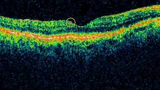
2/- if we have an abnormality in the OCT( GCC and RNFL) with normal clinical and perimetry , treatment or not ?
2/- Could you please have the generosity to make a video explaining the interpretation of Macular OCT and fluorescein angiography.
Best love and support and blessings from Algeria
As regards Q1 : yes you have to invert what you see with +90 D. Q2: OCT changes can occur in 1/3 of normal healthy persons with even small degree of myopia less than -4D. Diagnosis of glaucoma depends on +ve mutliple signs . so
for diagnosis of glaucoma you need o have more than OCT alone. The more the signs and symptoms the more one can be positive with the diagnosis
Q3 : I am sorry I am not a retina doctor to cover macular OCT or fluorescein agniography in practical way. hope you will fine some other source to help with this issue.
Hope to get a better voice quality for this excellent lecture
Sorry for the inconvenience. English subtitle is now shown, hope that will help.
thank you Doctor for these videos, I had read the myopia could cause the OCT for RNFL to be thinner, is it also the case for someone who's had myopia in the past but had LASIK, would it still cause the OCT RNFLY to be thinner? Thank you
Yes myopia in the past will result in possible RNFL thinning. LASIK has no effect on RFNL thickness.
@@adelabdelshafik thank you doctor, is your clinic only in egypt? so just to be clear, for myopia the possible RNFL thinning in OCT will still be the same after LASIK?
@@sorsara78 Yes, effect of myopia on RNFL thickness is not influenced by LASIK. Yes my clinic in Cairo, Egypt.
Dear Sir, can i ask, towards thr end of thr lecture, u say, upto 50%of -5DS myopes show errors of GCIPL and are fakse positive. So does is that basically mean, thst if we see a patient with apparent gcipl thinning, and fields normal and disc oct relatively normal, we should just monitor? And not treat?
The point to keep in mind is changes in OCT may occur in 1/3-1/4 of normal persons. So to diagnose glaucoma you need to have multiple different areas starting from family history, suggestive symptoms, abnormal angle configuration, suspicious disc, suspicious field changes, lower systemic BV, high myopia...... etc the more the abnormal factors the more the diagnosis is correct. so as you said, if not sure just keep pt under observation.
@@adelabdelshafik Thankyou...