Embryology of the Eye (Easy to Understand)

Vložit
- čas přidán 15. 05. 2020
- The development of the eyes explained in 15 minutes.
If you are completely new to embryology and you want to understand it quickly, this should be the first video you watch:
- • Introduction to Embryo...
Post any questions you have about the video below, I read all the comments:
--------------------------------
Recommended Text
--------------------------------
Easy Embryology is a book that is dedicated to the simplification of embryology. It is available at drminass.com/product/easyembr.... Contact Dr. Minass for more information.
----------------------------------------
Interact With Dr. Minass!
----------------------------------------
Website - www.drminass.com
Email - info@drminass.com
Patreon - / drminass
Facebook - / m1na55
Instagram - @m1.nass
Post - Address to:
Minass
Parcel Locker 10106 04448
59 Penshurst Street
Willoughby, NSW
Australia 2068
------------------------------------------------------------------------
SUMMARY OF THE VIDEO FOR YOUR NOTES
------------------------------------------------------------------------
Development of the eye is a little bit more complex than most of the organs already discussed, but as usual we will break it down simply so that we can make it easy to understand.
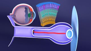
The eye is first noticed in week three of development. Just like the development of ears are marked with the appearance of an otic placode and then an otic vesicle, the eyes are marked by the appearance of the optic placode and vesicle on either side of the embryo. As the anterior and posterior neuropores close (see CNS section), the optic placodes will form into the optic vesicles. The optic placodes are developed from neural tube (ectoderm). The optic vesicles grow until they touch the ectoderm of the skin of the embryo. The lens’ then initiate their formation, at which point the vesicle evaginates to form the optic cup. The optic cup has two layers: an inner and an outer layer. The outer layer is the pigmented layer of the retina, and the inner layer is the neural layer. In week seven, the optic cup begins to form the pupil. The actual lens itself is developed from the surface ectoderm as a lens placode, and by week five detaches itself from the surface ectoderm and is in its position inside the optic cup.
The pars optica retinae is the outermost layer of the inner layer and will contain the rods and cones. Next to the pars optica retinae is the mantle layer which develops into the neurons of the eye (outer nuclear, inner nuclear, and ganglion layers). The axons of these nerves are on the surface and they join together to form the optic nerve (CNII). The most anterior portion of the inner layer has a one cell coating (pars ceca retinae) which will form both the pars iridica retinae (the inner layer of the iris) and the pars ciliaris retinae (becomes the ciliary body). The pars ciliaris retinae continually folds to give it its characteristic appearance and the ciliary muscle will eventually control the lens in response to light and other stimuli. The dilator pupillae and the sphincter of the eye form in the epithelial cells contained within the surface and the optic cup.
There is another inner and outer layer for you to remember now. By week six, the tissue that surrounds the developing eye will transform into an inner and outer layer. The inner layer will become a tissue resembling the pia mater, and the outer layer resembles the dura mater. The pia mater portion differentiates into the choroid, whereas the dura mater portion will become the sclera.
The anterior chamber forms when there is degeneration of the tissue that yet again forms an inner and outer layer. The inner layer is the iridopupillary membrane (which completely degenerates anterior to the lens) and the outer layer is the substantia propria. The cornea is made from the substantia propria, an epithelial layer from the ectoderm, and the epithelium shared with the anterior chamber. The posterior chamber is bordered by the iris and ciliary body. Both anterior and posterior chambers are in open communication with each other through the pupil, and both contain aqueous humor that continually circulates.
Between the retina and the lens, the vitreous body is formed by infiltrating tissue that initially surrounds the optic cup.


Easy Embryology (written by me) is the BEST textbook to use if you're infuriated with embryology. Get 20% off with code EASY20
Buy here -> drminass.com/product/easyembryology
He literally knows like where the confusion arises what a talent!!!
my embryology exam is literally in 2 days and I was struggling to find a proper eye development resources to study the eye from. You just saved me!! Thank you so much
Just in time 😎👌🏼
Lol, same, in 2 days lets gooo
Same mine is in 2hrs
Mine is in 4hrs
Mine is in 15hrs
This is just generally an exceptional explanation but the illustrations specifically are incredible - so clear and meticulous and aesthetically pleasing. I can't imagine erasing them!
I love your video - you're right, you make an extremely complex topic easy to understand and easy to remember with your animation (you being animated and involving yourself in the video, as well as likening yourself to an embryo lol!), and energy - the way you reiterate key points and go back to reminding us what the mesenchyme is for example, stressing those points - it was a 15 minute video that just flew by because it was so enjoyable! Bless you and people like you for helping the world :) thank you!
Thank you so much for these videos they're so helpful .... I'm watching every one of them as i study embryology and i will continue to watch every single one ❤❤
Thank you!!! I'm a student of optics with intentions for optometrics in the future. I have prepared for my presentation about human eyeball embryology from your video :) It was very helpful
dude literally you're saving me, i was looking at my embryology book and everything was like a foreign langauge. these videos make the material so easy to understand, thanks man!
It literally is in another language. No time to learn latin right
Dr Minass, you're exceptional and a blessing. So good to have teachers like you who know from start to finish the entire topic. You've really helped me revise during my ophthalmology rotation, thank you so much! Will always appreciate your effort.
Wow, thank you, best of luck
Thank you for all the effort you put into this video. Please continue making them. You are helping a lot 💓💓💓
Thanks will do!
Amazing!! Now I can watch my lecture without feeling like it’s a different language!
This is so beatifully explained, embryology isn't scary anymore! Thank you so much for your dedication, keep up with great work!
Thanks Milan!
Dr. Minass what a wonderful didactic lecture, simply fantastic and easy to understand. Congratulations once more!
Many thanks!
your way is really easy to understand . you deserve a lot of
appreciation . thank you.
You are most welcome. Thanks so much for watching
Thank you so much for the videos it has helped me a lot since we started studying online due to Covid19.. please do videos on other subjects especially neuro anatomy
I was literally pulling my hair trying to figure out which part of the eye originated where THANK YOU!
I legit tried to follow the color coordination of eye development figures.
You are a great teacher. You made this complex process clear and easy to remember. Thanks
My pleasure
So I have been kind of really hooked up and now it is so frustrating I can't read an embryology topic without watching one of your videos to get a head start. Thankyou so much ❤️
Thanks Javairia
Beautifully explaned thank you for saving my grade🙇🏼
Thanks for a good summary Doctor!
Thank you so much, sir, although the video had so many points I had to watch it repeatedly.
Fantastic video, really easy to follow and super helpful for my part 1 ophthalmology revision! Thanks!!!
Glad it was helpful!
Thank you so much Dr Minas
You hv explained this in such a great n simplified way..... I guess I can never be able to understand this good without ur help...
Your diagrams are way to informative and easy to understand...
Thanks a lot Dr. Minass
It's my pleasure. Thanks!
wow, appreciate you indeed. this is ever simplest video explaining all and every steps . thankyou
You are extremely welcome.
I love how you make them simple. Please keep it up ❤️
Thank you! 😊
Great work thanks dr minass
Nicely explained ... It cleared my concepts ⚡
Really helpful I was struggling to learn embryology. Thank you for all the videos on embryology
Happy to help!
Can't believe i can finally understand it , the diagrams are very clear and your process of thinking and showing every step in details yet it seems very simplified
May god bless you dr minass , ty alot 🙏💖
You are most welcome
Great explanation and great video, thanks a million
Thank you , amazingly explained video
You’re the best out the best!!
Please dont stop keep upload videos❤
You've helped me conceive embryology properly. Now I read for the extra details which seep in easily now. Thanks
Very welcome
Please upload more!! thank you so much
Great freaking video, all the other sources were so unclear as to which part of the eye came from where, but here, man, you're my hero. Big thanks!
Glad you enjoyed it! Thanks for watching
@@minass Literally had the exam today and guess what question comes up? Development of eye. Guess who got an A lol
Thank you very much you made it too easy to understand 💖
i nearly cried in happiness, thank you so much, your explaination is so easy to understand. you should be famous !
Haha, thank you for watching Nindita
You seams to like so much that you made me like this too. Thanks !
Thank you so much sir ! You’re my lifesaver to my upcoming histology and embryology exam, may you also make an ear and skin development video? Thanks for saving many students out here :)!
Sure I will. This week
Thank you very much it's very helpful
very vividly explained sir with crystal clear diagrams. thank u!!!
You are welcome
Thank you 💕 for great effort 😊
Thankyou Sir for this amazing video, was struggling since so long to understand the basic embryology of eye. Really helped me understand :)
Glad it helped!
great tutorial video😊
Thank you so much sir for all these awesome videos. I'm inspired by your understanding of these intricate concepts and also by your ability to teach them. God bless you.
Thanks for watching!
Thats awasome thank you
You are a master ❤
This was amazing!!! Saving my grade and helping me actually understand!!
Thanks for watching!
Hello, thanks a lot for the brief description.
I would like to know about iris epithelium and ciliary process epithelium, are they developed from the inner layer of the optic cup?
Thank you so much sir ... Because of you I understand the embryology of eye ...
Glad to hear that!
THANK YOU , THAT WAS REALLY HELPFUL
YOU'RE WELCOME!!
Thank you 🙏🏽
Thank you!
Great work Dr. Minass
It's my pleasure
Amazingly clear .. Thank you!
Great to hear!
Dr. Minass have got a doubt in this video and in general about embryology here when you switched from the diagram from when the lens placode invaginates and the optic stalk becomes double walled to the diagram where you show the optic stalk I couldn't see the continuation properly. Idk if it just me but in general when the axes change in embryology it confuses the heck out of me. Any ideas on how to make sense of this ?
Sir you are great as a teacher. You shouldn't have discontinued teaching. Where were you, we were waiting ?
It’s like finding a rare Pokémon
Just awesome 😀
many thanks😊
That was great..i'm so grateful for you 🙏
I'm grateful that you watched the video and it was helpful. Enjoy
you are amazing!!!
Got 1 subscriber sir,you nailed it.❤️❤️
Great explanation, thank you!!!
Quite welcome
Thank you very much sir
thanxx a lot sir! helped real good :D
Thank you so much for this wonderful video.🤗🤗
Most welcome 😊
Thanks this helped😊
you always save my life in embryo !!! my exam is in 2 days !!! thanks
Another life saved 👌🏼😎
Thank you for this awesome video !
Glad you liked it!
Thank you! Easy to understand and the pictures are just perfect!!
Thx again!
So useful, thank you !!! perfect timing for my senses test ;) !
Keep up!
Thanks for watching Pilar!
Hi am Malon from Kenya and you
@@malonkipngenoh2856 hey what's up, im from Portugal !! dope
Thank you sir❤
It's very nice..
Love from Pakistan ❤️ it was really helpful
Explained better than a PhD at my medical school... Thanks
Wow you simplified the work for me
Thanks for watching. I try to make everything easy for students!
Dr. Minass love you sir
🥰
U r the best
Thanks, nicely explained.
Glad it was helpful!
For a person who was born with one eye not finally formed, opened preauricular pits and nipples retraction, would it be possible to guess in which week of gestation complications were presented?
Any situation by which the lens and cornea gets blood flow again? Perhaps allowing future gene therapies to regenerate them.
Really great video ! Do you think it's possible to make a vid about aortic arches to complete your series about pharyngeal development ??
Great idea. Will do soon 👌🏼
Thank you so much I love your videos
Thanks Timi 👌
I watched your video when I was in my Year 2 struggling with embryology. And now you help me again. Thanks a lot Dr. Minass !!!
Its my pleasure Leo. Keep up the good work!
Thank you, made it easier to read KLM.
Soon, you won't need it hehe
Thank you so much DrMinass.
Can you make a video specifically on birth defects. I know it would require at least 2 videos of 15 mins each but still if you could do it it would be great.
Anyways have a great day and may god bless us all.
Sure that’s a good idea 👌🏼
Hi Dr Minass. I've heard that about a million nerve cells from each eye grow and connect in a junction which in turn connects to the brain. I've also heard that each nerve cell from one eye must connect to the correct nerve cell from the other eye. In other words the nerve cells from each eye are paired in specific pairs, and not just random connections. Would you be able to explain this more clearly for me?
Thank you ! very helpful
Glad it was helpful!
omg thank you so much please do one on the ear :)
I will!
This is really amazing pls upload more videos
Fun fact I am not even in medical college yet but it's really amazing 👍🏼👍🏼👍🏼🤟🤟👍🏼
Thanks sir for your help through this video 👍💖
Always welcome
Thank you, It’s really useful . Besides, can you make a video about the embryology of ears?
Yes, soon
Thank you sir. This helps a lot :)
You are most welcome
i am confused about the conjunctiva, lacrimal apparatus ( lacrimal gland, sac, nasolacrimal duct and canaliculi)
Very much helpful .... Thank you Sir ..
All the best
Thank you sir🥰🥰🥰
Have a great day. you make our days great. so I wish same for you sir
Making your day great was the ultimate goal of this video 👍
awesome video. thanks alot.
Glad you liked it!