obviously this video is basic and for beginners. but for those wanting to obtain difficult lines using the US, i suggest tilting the probe away from puncture site to get the best reflection and best view of the shaft. once identified the shaft > slide the probe away from puncture site keeping the visuals on the shaft that will soon exhibit a double dot opacity which signifies the edge of the tip. and then advancing the needle towards the vascular anechogenicity. once pierced, shallow angle is opted, and backflow may be noted but do not under any circumstance slide in the sheath yet; ignore the backflow, continue to advance the needle further within the veascular anechogenicity keeping the tip in the center (to avoid puncturing posterior wall of the vessel) ; eyes all the time on ultrasound. utilize a blinking method, where once the vascular anechogenicity becomes dark, advance the tip until visualized in the center and then slide the probe further again until the tip is no longer visualized and then repeat. when a good amount of needle is within the vessel, u can opt to double confirm using longitudinal views or simply slide the sheath within the vessel. difficult veins require careful strategy involving pythagoras calculation, ascertaining the correct trajectory and depth of the vessel and puncturing the correct length from US probe.
After the Tegaderm is placed on the probe and smoothed with a bare hand or even a gloved hand I don't believe it's semi sterile any longer. Can you clarify? And if it's not could you leave the Tegaderm off?
Basilic is actually where we prefer to place PICC lines, cephalic and brachial are lesser options. Use caution when trying the brachial veins, they are very close to the brachial artery and median nerve! The safest place to put an USG IV is the cephalic, since that is father away from the nerve, and it is not the preferred site for a PICC line.


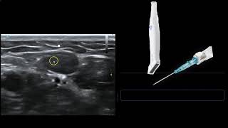


obviously this video is basic and for beginners. but for those wanting to obtain difficult lines using the US, i suggest tilting the probe away from puncture site to get the best reflection and best view of the shaft. once identified the shaft > slide the probe away from puncture site keeping the visuals on the shaft that will soon exhibit a double dot opacity which signifies the edge of the tip. and then advancing the needle towards the vascular anechogenicity.
once pierced, shallow angle is opted, and backflow may be noted but do not under any circumstance slide in the sheath yet; ignore the backflow, continue to advance the needle further within the veascular anechogenicity keeping the tip in the center (to avoid puncturing posterior wall of the vessel) ; eyes all the time on ultrasound. utilize a blinking method, where once the vascular anechogenicity becomes dark, advance the tip until visualized in the center and then slide the probe further again until the tip is no longer visualized and then repeat.
when a good amount of needle is within the vessel, u can opt to double confirm using longitudinal views or simply slide the sheath within the vessel.
difficult veins require careful strategy involving pythagoras calculation, ascertaining the correct trajectory and depth of the vessel and puncturing the correct length from US probe.
wow awesome video, thanks!
Also..you can damage prove over time by not using a bit of lube under Tegaderm
After the Tegaderm is placed on the probe and smoothed with a bare hand or even a gloved hand I don't believe it's semi sterile any longer. Can you clarify? And if it's not could you leave the Tegaderm off?
Was thinking the same. A better method would be to cover the probe with a sterile glove with some jelly inside.
It’s not sterile anymore, which defeats the whole purpose of covering the probe.
Basilic is actually where we prefer to place PICC lines, cephalic and brachial are lesser options. Use caution when trying the brachial veins, they are very close to the brachial artery and median nerve! The safest place to put an USG IV is the cephalic, since that is father away from the nerve, and it is not the preferred site for a PICC line.
MediAN nerve.
Shut up
5:37 cephalic vein is more pulsatile even with torniquet on !!! How to clarify and make sure it is the vein and not artery !!!!
That is not pulsating. It is actually being compressed and released to confirm it is a vein and rule out thrombus
Hitting the vein just easy but previnting infiltrate is hard
i liked how fast it was but it felt like a Tac going in my skin other than an IV to me
I find 18g are easier to see the tip