Histology of cervix - Shotgun Histology

Vložit
- čas přidán 24. 05. 2019
- 📌 𝐅𝐨𝐥𝐥𝐨𝐰 𝐨𝐧 𝐈𝐧𝐬𝐭𝐚𝐠𝐫𝐚𝐦:- / drgbhanuprakash
📌𝗝𝗼𝗶𝗻 𝗢𝘂𝗿 𝗧𝗲𝗹𝗲𝗴𝗿𝗮𝗺 𝗖𝗵𝗮𝗻𝗻𝗲𝗹 𝗛𝗲𝗿𝗲:- t.me/bhanuprakashdr
📌𝗦𝘂𝗯𝘀𝗰𝗿𝗶𝗯𝗲 𝗧𝗼 𝗠𝘆 𝗠𝗮𝗶𝗹𝗶𝗻𝗴 𝗟𝗶𝘀𝘁:- linktr.ee/DrGBhanuprakash
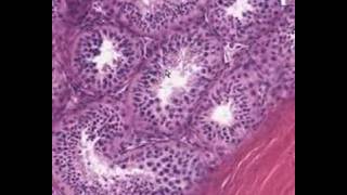
Histology of cervix
Ectocervix
-----------------
Stratified non keratinizing squamous epithelium
Basal cells: deepest layer; dense nuclear chromatin, uniform oval nuclei oriented perpendicular to the basement membrane, scant cytoplasm
Parabasal cells: located just above the basal cell layer; slightly more cytoplasm than basal cells; maybe multiple cell layers thick
Intermediate cells: abundant cytoplasm which may be pink or clear due to glycogen accumulation
Superficial cells: small, round nuclei; abundant pink or clear cytoplasm; cells flatten and are oriented parallel to the basement membrane
Hormone responsive
Superficial cells predominate in the early cycle due to estrogen
Intermediate cells predominate in late-cycle due to progestins
Loss of intermediate and superficial cells (atrophy) occurs postmenopause
Rare melanocytes, Langerhans cells, and endocrine cells have been identified.
Endocervix
------------------
Single layer of mucinous columnar cells with dense, uniform, oval, basally oriented nuclei and apical mucin
Mucin has a pale bluish appearance in H&E preparations; with PAS-Alcian blue stain, apical mucin stains intense blue / purple (due to the presence of acid type mucin)
Ciliated cells can be found (usually in the context of tuboendometrioid metaplasia)
Inconspicuous underlying reserve cell layer
Forms infoldings, clefts and glands of variable shape
Transformation zone
----------------------------------
Metaplastic cells: formed by endocervical reserve cells differentiating toward squamous lineage
Located at transition between glandular and squamous epithelia
Similar appearance to parabasal cells with relatively scant cytoplasm and dense nuclei
Endocervical epithelium may overlie metaplastic cells
Variable nonspecific inflammatory infiltrate consisting of lymphocytes, plasma cells and even neutrophils is common and is not necessarily associated with infection
Cervical stroma
--------------------------
Mostly fibrous tissue with some haphazard smooth muscle fibers
Blood vessels often numerous and prominent
Mesonephric rests / remnants
-------------------------------------------------
Remnants of involuted embryologic mesonephric (Wolffian) ducts
Present in lateral cervical wall in ~33% of women
Microscopic clusters of tubules lined by single layer of cuboidal cells with eosinophilic luminal secretions
#histologyofcervix #cervixhistology #histologycervix #shotgunhistologycervix #shotgunhistology


Excellent explanation! The arrows and dashed lines were quite helpful to understand the details. This is indeed the nicest cervix.
How to differentiate between squamous metaplasia and stratified squamous?
Thank you this was helpful
Glad it helped
Cervix has no serosa or adventitia ?
Thank you
🤝🤝🤝
Credit goes to Washington deceit
yes we have taken written permission