Thank you for this! I am a student at a different community college but this was exactly what I needed to know for my practical tomorrow.I am very confident that I will do well now! :)
You are the best. The absolute best. No Joke. I was trying to learn the eye with pictures in textbook but the correct way to learn it is using a model like you showed here. Thanks!
I have to thank you. I just looked into this in more detail. I was confusing the segments of the eye with the chambers. Both of those compartments are part of the anterior segment. Thanks man.
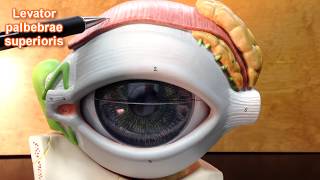
Here's your list with short explanations of what each part is. I hope it helps :) ora serrate -- the area between the retina and the cilliary body. cilliary process -- part of the cilliary body that produces aqueous humor. cilliary muscle -- smooth muscle in the cilliary body, its contraction controls the focusing action of the lens (accommodation).
Thx!! This helped me out for my test coming up Wednesday! I liked both of your eye videos. I don't go to school, or live in your state, but you should keep posting these videos:)
I've watched 20 videos on the eye today and I think you have the best. Nice and simple and easy to understand. .. side note I thought you were black lol your voice n hands smh. Good job thanks
Circular muscle and radial muscles are part of the iris. On this model you can't really see them. The circular muscle is smooth muscle within the center most part of the iris (close to the pupil), when it contracts the iris closes making the pupil smaller (less light enters the eye). The radial muscles are also smooth muscle. These muscles radiate from the pupil kind of like spokes on a bicycle. When the radial muscles contract they cause the pupil get larger (more light enters the eye).
The model I'm using here does not have suspensory ligaments (at least not well shown) so I consider that area simply the ciliary body. In graduate school I used to dissect human eyes and I never saw the suspensory ligaments, I think because they are very small fibers. The next time I dissect fresh cow eyes I'll try to visualize the suspensory ligaments again. If it works I'll see if I can post some images or a video.
Yes, the ciliary muscle is part of the ciliary body. It is a circular smooth muscle that controls the shape of the lens. When the ciliary muscle is relaxed the lens is more flattened-- this is the position for distance vision. When the ciliary muscle contracts the lens become thicker-- this is the shape needed to see close objects.
This video has been very helpful.I watched a lot of videos today on the eye and yours is the best. Thanks a lot! But can you please talk about the tunics. Thanks all the same.
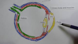
Thank, and I'm glad the videos are helping. "Tunic" is a fancy way to say "layer." The sclera or "white" of the eye is the outer tunic (fibrous tunic). The choroid layer is the middle tunic, it is highly pigmented and also rich with blood vessels (vascular tunic). The retina is the innermost layer (sensory tunic). Let me know if you have any more questions :)
Hi, I have an anatomy test on the eye on Friday, we only had 20 minutes of lab! so I have been researching like crazy these certain parts because my book does not show, I am having trouble with the ora serrate, cilliary process, and the cilliary muscle, I think they are all in the retina? it is hard to tell though, also the rods would we be able to actually "see" the rods on our test, also the conjunctiva? thanks a lot ! :)
They are located superolaterally to the eyes (above and to the outside of the eyes). If you do a search for images of lacrimal gland you will quickly find images of what I mean :)
Very helpful except I'm a little confused about the compartments, my text book says that what you are calling the posterior compartment is the vitreous chamber, and that the posterior chamber is between the iris and the lens, the anterior chamber is between the iris and the cornea.
@@renhartung yes. I also confirmed with other professors and looked it up in the text book Human Anatomy and Physiology page 548 on special senses by E. Amerman
Yes I do. Click on my name in any of these messages and you will be taken to my channel, there you can see my playlists. Look in the playlist called "Cardiovascular System"
Glaucoma is a disorder causing damage to the optic nerve... the most common type involved increased pressure inside of the eye. There is a constant flow of fluid into and out of the eyes, if the outflow is not functioning properly pressure builds. That pressure compresses the optic nerve against the sides of its exit/entry point (the lamina cribrosa) at the back of the eye.


The best youtube Anatomy and Physiology instructor here. Thank you for all you do. You are my go to for help with the models.
I really really understand the eye now, better than the way my A&P professor taught it. Thanks a lot! God bless you!
I have my lab test tomorrow and feel as confident as ever after watching your video over and over again. Thank you for the good work :)
Woooww!!! This is the best video out here! Thank you professor Hartung for easing my pain! :)
Thank you for this! I am a student at a different community college but this was exactly what I needed to know for my practical tomorrow.I am very confident that I will do well now! :)
You are the best. The absolute best. No Joke. I was trying to learn the eye with pictures in textbook but the correct way to learn it is using a model like you showed here. Thanks!
Best video on CZcams describing the eye!
This is the best eye presentation ever. I'm sure I won't miss any part on my lab practical.
Very good explanation and very great model as well
i wish my professor is as clear as Dr. Ren Allen. Thank you so much sir.
I wish my professor were as clear as you. And had visual diagrams. Thanks! This will help me pass my lab final!
Thank you for this wonderful video! I have a lab exam on Monday, and this helped me so much.
Thank you so much!
It was the model I needed & also had further explanations that my Professor did not provide!
such an excellent video for lab! More students need to watch these especially with virtual learning.
thank you so much for this. I am at a different college and was SO helpful!
Cheers, this has helped me so much! Subbed!
Thanks! you explained everything thoroughly, just made ophthalmology super easy!
I have to thank you. I just looked into this in more detail. I was confusing the segments of the eye with the chambers. Both of those compartments are part of the anterior segment. Thanks man.
I WILL pass my exam because of your video and a lot of hard work...thanks
thank you! this video helped me understand it a lot better.
Here's your list with short explanations of what each part is. I hope it helps :)
ora serrate -- the area between the retina and the cilliary body.
cilliary process -- part of the cilliary body that produces aqueous humor.
cilliary muscle -- smooth muscle in the cilliary body, its contraction controls the focusing action of the lens (accommodation).
Thx!! This helped me out for my test coming up Wednesday! I liked both of your eye videos. I don't go to school, or live in your state, but you should keep posting these videos:)
Thank you so much this video has been a great help, i have my lab practical thursday
This video was so clear and helpful thank you so much !!!
Amazing video! Explains a lot
I've watched 20 videos on the eye today and I think you have the best. Nice and simple and easy to understand. .. side note I thought you were black lol your voice n hands smh. Good job thanks
thank you very much professor!! this was really helpful
THANK YOU DOCTOR HARTUNG. U ROCK:)
Best video i've watched, very clear and easy to understand!
😉
Thanks! This video was really helpful!!!
Circular muscle and radial muscles are part of the iris. On this model you can't really see them. The circular muscle is smooth muscle within the center most part of the iris (close to the pupil), when it contracts the iris closes making the pupil smaller (less light enters the eye). The radial muscles are also smooth muscle. These muscles radiate from the pupil kind of like spokes on a bicycle. When the radial muscles contract they cause the pupil get larger (more light enters the eye).
Love this video ...very helpful
Thank you Sir Believe that your lecture is excellent and I enjoy them and many problems are solved
Great job!
Thank you for posting this
Yes, the larger area is called the macula. At the center of the macula is the fovea centralis :)
Cheers, the dark circle lateral to optic disk is also called the macula?
Best video about eye model
Nice presentation
Question: what you had label #36 aren't those the suspensory ligaments and the red fan like structure the ciliary body?
The model I'm using here does not have suspensory ligaments (at least not well shown) so I consider that area simply the ciliary body. In graduate school I used to dissect human eyes and I never saw the suspensory ligaments, I think because they are very small fibers. The next time I dissect fresh cow eyes I'll try to visualize the suspensory ligaments again. If it works I'll see if I can post some images or a video.
thank you so much! :D
Yes, the ciliary muscle is part of the ciliary body. It is a circular smooth muscle that controls the shape of the lens. When the ciliary muscle is relaxed the lens is more flattened-- this is the position for distance vision. When the ciliary muscle contracts the lens become thicker-- this is the shape needed to see close objects.
Awesome this was so much better than the dvd copy St. Philips gave us!
Plus you sound like the movie star Bradley Cooper lol
Excellent
Thanks!
I really understand, good tutorial
thanks do you have heart models on your channel ?
Thank you so much 🙂
I. LOVE. THIS. VIDEO!!!!!!!!!
This video has been very helpful.I watched a lot of videos today on the eye and yours is the best. Thanks a lot! But can you please talk about the tunics. Thanks all the same.
Thank, and I'm glad the videos are helping.
"Tunic" is a fancy way to say "layer." The sclera or "white" of the eye is the outer tunic (fibrous tunic). The choroid layer is the middle tunic, it is highly pigmented and also rich with blood vessels (vascular tunic). The retina is the innermost layer (sensory tunic). Let me know if you have any more questions :)
thanks for this
thanks
THANK YOU SO MUCH THIS WAYS NO HELP
Hi, I have an anatomy test on the eye on Friday, we only had 20 minutes of lab! so I have been researching like crazy these certain parts because my book does not show, I am having trouble with the ora serrate, cilliary process, and the cilliary muscle, I think they are all in the retina? it is hard to tell though, also the rods would we be able to actually "see" the rods on our test, also the conjunctiva? thanks a lot ! :)
How did you make it
They are located superolaterally to the eyes (above and to the outside of the eyes). If you do a search for images of lacrimal gland you will quickly find images of what I mean :)
thankyou
excellent
Are the ciliary muscles inside the ciliary body?
i like it. thank u sir
good video
Thanks !!!! :)
do you know where the lacrimal gland is located ?
What is the vein that lies next to the central retinal artery air?
Central retinal vein drain to superior ophthalmic vein then to cavernous sinus
none of these videos have the caruncles? and the canthus??
The central vein lies right next to the central artery :)
Very helpful except I'm a little confused about the compartments, my text book says that what you are calling the posterior compartment is the vitreous chamber, and that the posterior chamber is between the iris and the lens, the anterior chamber is between the iris and the cornea.
also where is the macula lutea?
Number 36 are the suspensory ligaments and the muscle underneath is ciliary body
So, to you the white lines on the model represent zonular fibers (suspensory ligaments as you say). That makes sense to me.
@@renhartung yes. I also confirmed with other professors and looked it up in the text book Human Anatomy and Physiology page 548 on special senses by E. Amerman
my right ear says thank u ;D
where is circular muscle and radial muscles?
Yes I do. Click on my name in any of these messages and you will be taken to my channel, there you can see my playlists. Look in the playlist called "Cardiovascular System"
Please can explain how glaucoma occurs or happens
Glaucoma is a disorder causing damage to the optic nerve... the most common type involved increased pressure inside of the eye. There is a constant flow of fluid into and out of the eyes, if the outflow is not functioning properly pressure builds. That pressure compresses the optic nerve against the sides of its exit/entry point (the lamina cribrosa) at the back of the eye.
Thanks a million
lol did he fail to mention the retina??
Ora serrata