Thigh cross sectional anatomy - Muscular Compartments - Adductor Canal

Vložit
- čas přidán 22. 03. 2023
- #thigh #anatomy #sciatica
Link for Donations paypal.me/studentlamedicina?l...
/ anatomy.knowledge
We will draw a diagram of a cross section through the middle third of the thigh. First we will indicate the femur which posterior presents a ridge called linea aspera. Muscles of the thigh are covered by a strong fascia called fascia lata (or the deep fascia of thigh). Lying on it is the superficial fascia of thigh composed of areolar tissue. And most superficial is of course the skin of thigh.
Now we are in a position to indicate the four sides of the thigh: L, M, A and P.
Laterally the fascia lata is thickend by the presence of the iliotibial tract.
Medially and superficial to the fascia lata we can observe a cross section through the great saphenous vein.
From the linea aspera to the fascia lata are diverging three intermuscular septa.
The lateral intermuscular septum, posterior intermuscular septum and medial intermuscular septum. These septa divide the thigh into three muscular compartments.
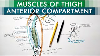
The anterior compartment contains the following muscles: Originating from the anterior and lateral and medial sides of the shaft of femur is the vastus intermedius muscle. Lateral and superficial to it is the vastus lateralis. Medial and superficial to vastus intermedius is the vastus medialis. In front of vastus intermedius is the rectus femoris. We cand also observe medially and just under the facia lata, the Sartorius muscle.
In the medial compartment or the adductor compartment we can observe on this cross section , the adductor longus muscle which lies just behind the medial intermuscular septum. The adductor magnus muscle ocupyies most of the compartment, and medially is the gracilis muscle. Deep to these muscles are the deep femoral vessels.
In the posterior compartment we draw laterally the biceps femoris muscle which is composed of long head and short head. Medially to this muscle are the semitendinosus and semimembranosus muscles. Deep to these muscles we observe the siatic nerve which supplies them.
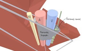
We are now in a postion to discuss about the adductor canal which is also called the subsartorial canal or Hunter’s canal. This canal is located in the medial side of the anterior compartement and on a cross section is triangular in shape thus having three borders. Anterior border is represented by the vastus medialis, the posterior border is the medial intermuscular septum and the adductor longus muscle. Lateral border is the Sartorius muscle which lies on the vasoadductor membrane.
The adductor canal contains the femoral artery and branching from it the descending genicular artery, the femoral vein and two nerves: branch to vastus medialis and the saphenous nerve.


very neat and clear
I got distinction and CLS topper by listening Ur easy explanation tnqu so much❤
god bless you for taking so much efforts to teach
Awesome
Keep doing sir
Best wishes ❤️
Superb
Sir, please make
videos on other parasympathetic ganglions such as otic,pterygopalatine and mandibular..
you are literally better then many professors i know, love your content, you make anatomy fun, i dont understand how you have less followers when literally everybody loves your content
Thnx ❤
❤
Stomach bed plz..... From Bangladesh 🥰
Sir,Make sketch video about superior vena cava
3:39
🫡
Stop making anatomy so easy