Hernia Ultrasound Reporting | Direct/Indirect Inguinal/Epigastric/Femoral Hernia USG Reports

Vložit
- čas přidán 4. 07. 2024
- Hernia Ultrasound Reporting | Direct/Indirect Inguinal/Epigastric/Femoral Hernia USG Reports
*Cases
Intro - 0:00
Epigastric Hernia - 0:10
Spigelian Hernia - 3:24
Umbilical Hernia - 6:18
Hypogastric Linea Alba Hernia - 8:31
Incisional Hernia - 11:07
Richter Hernia - 14:01
Direct Inguinal Hernia - 16:36
Indirect Inguinal Hernia - 19:24
Pantaloon Hernia - 22:03
Amyand Hernia - 25:18
Scrotal Hernia - 27:46
Femoral Hernia - 30:07


Hernia Ultrasound Normal Vs Abnormal Images | Direct/Indirect Inguinal/Epigastric/Femoral Hernia USG: czcams.com/video/FCeS3Kx_vKE/video.html
You are the best thank you very much
Thank you so much for watching!
Tnx Dr Sam. Nice 👍 video
Most Welcome!
BEST PRESENTATION
Thank you very much for watching!
Excellent
Thanks for watching!
Very informative 🧐
Thanks for watching!
Nice and very informative as always...
Thanks for watching!
@@DrSamsImagingLibrary keep up with the good work sir .
ver informative
Thanks for watching!
Thanks so much sir....for sharing your knowledge with us,my humble question is how to differentiate an inguinal hernia from a scrotal hernia sonographically
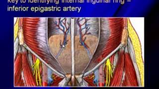
Most Welcome and thanks for watching! An inguinal hernia is identified as a protrusion through the inguinal canal, which is located above the inguinal ligament and often medial to the inferior epigastric vessels. A scrotal hernia is identified as a herniation that extends into the scrotum. It can originate as an inguinal hernia but has descended further down into the scrotum.
Sir which test detects hernia.i have a doubt which test should I go first ultrasound abdomen or ct abdomen??
You should have an ultrasound first. It can detect hernias.
Hello..
I am 19 weeks pregnant.
Tiffa scan lo FL value 22 mm 16 weeks 6days.
Bilateral short femour length with bowing deformity..
Bilateral short humerus with radius.
Is it normal.. Plz sir am so worried abt it
Hello. I'm sorry to hear about your concern. This may suggest a skeletal dysplasia. The findings are abnormal
@@DrSamsImagingLibrary thank you..