MSK Ultrasound Evaluation: Differentiation of Tendinopathy vs Tear in the Rotator Cuff

Vložit
- čas přidán 27. 07. 2024
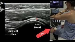
- In this video, a collaborative project between AIUM and APTA, Colin Rigney PT, DPT, OCS, RMSK, explains the terms high-grade and low-grade, with respect to rotator cuff tendon pathology; shares probe positioning, patient positioning, and image acquisition with respect to rotator cuff tendon evaluation; teaches (via MSKUS imaging) how to establish a working algorithm to help determine/define a partial thickness tendon tear versus tendinopathy; and shows tendon shape and echotexture(s) that represent high-grade pathology associated with partial and full-thickness rotator cuff tears.
Interested viewers may be able to earn CME credit. If available, it is located here: learn.aium.org/products/msk-u...
Original air date: 9/21/2018


Very helpful lecture. Thank you.
very instructive . thanks.
Great lecture!
Very informative
love you sir , great lecture
Thank you sir.
شكرا سيدتي
When youre showing the tendinopathies you should show where the ultrasound head is positioned.
👌🌹8:56
*為SS 肉-腱交界的#肌肉組織
9:59 SS 腱著處=footprint約1.5cm
10:21 SS腱短軸觀
10:55 IS 腱 11:31
12:05 SubScap腱
🌹20:13 腱病變Tendinopathy
🌹26:58 Chr tear可見GT的皮質骨表面不平整。
未完待續
great