Introduction to Peritoneum

Vložit
- čas přidán 17. 11. 2020
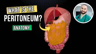
- Anatomy of peritoneum is a challenging topic yet it is important to understand the organization of the abdominal organs and peritoneal cavity.
- Parietal peritoneum lines the abdominal walls and diaphragm. It is supplied by same nerves and blood vessels of the abdominal walls and diaphragm (intercostal and phrenic nerveves and vesssles.
- Visceral peritoneum, represents the serosa layer of the abdominal organs and receives the same nerve and blood supply as related organs.
- Peritoneal cavity is divided into large and lesser sac.
- Lesser sac is located behind the liver, lesser omentum, stomach and anterior wall of greater omentum. Bounded superiorly by diaphragm and closed inferiorly by the reflection of greater omentum.
Both lesser and greater sacs are communicating through the epiploic foramen that is bounded anteriorly by portal triade (hepatic artery, common bile duct and portal vein) and posteriorly by inferior vena cava.
- Important peritoneal reflections and folds include and not limited to
1. Falciform ligament
2. lesser omentum
3. greater omentum
4. transverse mesocolon
5. mesentry
6. segmoid mesocolon
7. mesoappendix
8. gastrosplenic ligament
9. spleno-renal ligament
10. broad ligament of uterus
- These folds and ligaments carry blood, nerve supply and lymphatic to the organs.
- Organs can be completely, partially covered or retroperitoneal.
- Examples of completely covered organs: Liver, stomach, spleen, small intestine, appendix, Transverse colon, segmoid colon
- Examples of partially covered organs ascending and descending colon
- Examples of retroperitoneal organs: most of duodenum, pancrease and kidneys.
- The peritoneal cavity above transverse colon is divided into right and left subphrenic spaces by falciform ligamens. These spaces continue down to paracolic gutter in the right and left sides and could be a place for accumulation of fluid/blood or pus in case of infection, ascites, or heamorrahge.
- Perforated organs can lead to peritonitis e.g perforated gastric ulcer leads to leakage of gastric juice to lesser sac that may cause peritonitis, pancreatitis or even injury of splenic artery.
- Excess production of peritoneal fluid is called Ascites and could occure as a complication of some diseases such as liver cirrhosis, or congestive heart failure.
- Peritonitis can also lead to adhesion between the parietal and visceral layers. - Jak na to + styl


Great video
Thanks 👍🏽
Amazing
I enjoy your videos....these are so effective!.
Can you make video on heart...!!
الفاينل قرب يا بروف في اي اخبار على ال body walls🙏🙏🙏
I'm so sorry, i have been extremly busy. I promise once i have sometimes i will do it immediately.
Best luck!
@@EasyAnatomy جدع من يومك يا بروف
Dear everyone, this is a link to kahoot interactive questions that briefly cover the anatomy of digestive system and peritoneum. Copy the link to a new browser and answer the questions.
kahoot.it/challenge/04182796?challenge-id=c2a3c554-de6f-4efe-a708-c61a22d428c8_1605770541534